Difference between revisions of "Documentation/Nightly/Announcements"
| Line 136: | Line 136: | ||
Image:CMRTK-logo.png|[[Documentation/{{documentation/version}}/Extensions/CARMA|Cardiac MRI Toolkit]] to analyze cardiac LGE-MRI images. {{updated}} | Image:CMRTK-logo.png|[[Documentation/{{documentation/version}}/Extensions/CARMA|Cardiac MRI Toolkit]] to analyze cardiac LGE-MRI images. {{updated}} | ||
| − | Image:CarreraSliceEffect.png|[[Documentation/{{documentation/version}}/ | + | Image:CarreraSliceEffect.png|[[Documentation/{{documentation/version}}/Modules/CarreraSliceInteractiveSegmenter|CarreraSliceInteractiveSegmenter]] to interactively segment in 3D {{updated}} |
| − | Image:I2M_logo.jpg|[[Documentation/{{documentation/version}}/Extensions/CBC_3D_I2MConversion|CBC 3D (CRTC's BCC & Compression) I2M (Image-To-Mesh) Conversion]] for Image Guided Therapy. The extension encapsulates two CLI modules: (1) Body Centric Cubic (BCC) Mesh Generation | + | Image:I2M_logo.jpg|[[Documentation/{{documentation/version}}/Extensions/CBC_3D_I2MConversion|CBC 3D (CRTC's BCC & Compression) I2M (Image-To-Mesh) Conversion]] for Image Guided Therapy. The extension encapsulates two CLI modules: (1) Body Centric Cubic (BCC) Mesh Generation. (2) Mesh Compression (MC). {{new}} |
Image:ChangeTracker logo.png|[[Documentation/{{documentation/version}}/Extensions/ChangeTracker|Change Tracker]] for quantification of the subtle changes in pathology.{{updated}} | Image:ChangeTracker logo.png|[[Documentation/{{documentation/version}}/Extensions/ChangeTracker|Change Tracker]] for quantification of the subtle changes in pathology.{{updated}} | ||
| Line 156: | Line 156: | ||
Image:DTIProcess-mj.png|[[Documentation/{{documentation/version}}/Extensions/DTIProcess| DTI Process]].{{updated}} | Image:DTIProcess-mj.png|[[Documentation/{{documentation/version}}/Extensions/DTIProcess| DTI Process]].{{updated}} | ||
| − | Image:FastGrowCutEffect.png|[[Documentation/{{documentation/version}}/ | + | Image:FastGrowCutEffect.png|[[Documentation/{{documentation/version}}/Modules/FastGrowCut|FastGrowCut]] is an editor effect providing a fast implementation of the GrowCut method that supports multi-label segmentations {{updated}} |
| − | Image:FinslerTractography.png|[http://www.nitrc.org/projects/finslertract/|FinslerTractography FinslerTractography] implements the Finsler tractography method with HARDI data as described by J. Melonakos et al., Finsler Active Contours, IEEE Trans PAMI, 30:412-423, 2008 | + | Image:FinslerTractography.png|[http://www.nitrc.org/projects/finslertract/|FinslerTractography FinslerTractography] implements the Finsler tractography method with HARDI data as described by J. Melonakos et al., Finsler Active Contours, IEEE Trans PAMI, 30:412-423, 2008. {{new}} |
| − | Image:GelDosimetry_Logo_128x128.png|[[Documentation/{{documentation/version}}/ | + | Image:GelDosimetry_Logo_128x128.png|[[Documentation/{{documentation/version}}/Modules/GelDosimetry|GelDosimetryAnalysis]] is a [[Documentation/Nightly/Developers/Slicelets|Slicelet]] covering the gel dosimetry analysis workflow used in commissioning new radiation techniques. {{updated}} |
Image:GyroGuide.png|[[Documentation/{{documentation/version}}/Extensions/GyroGuide|GyroGuide]] determines the trajectory angle and depth of puncture path, and transmits the calculated information to the probe for real time navigation. {{new}} | Image:GyroGuide.png|[[Documentation/{{documentation/version}}/Extensions/GyroGuide|GyroGuide]] determines the trajectory angle and depth of puncture path, and transmits the calculated information to the probe for real time navigation. {{new}} | ||
| Line 166: | Line 166: | ||
Image:IASEM.png|[[Documentation/{{documentation/version}}/Extensions/IASEM| IASEM]] to segmentation and process of IASEM Electron Microscopy images.{{updated}} | Image:IASEM.png|[[Documentation/{{documentation/version}}/Extensions/IASEM| IASEM]] to segmentation and process of IASEM Electron Microscopy images.{{updated}} | ||
| − | Image:IGynePyIcon.png|[https://github.com/gpernelle/iGyne iGyne] is an open source software for MR-Guided Interstitial Gynecologic Brachytherapy | + | Image:IGynePyIcon.png|[https://github.com/gpernelle/iGyne iGyne] is an open source software for MR-Guided Interstitial Gynecologic Brachytherapy. {{new}} |
| − | Image:IntensitySegmenterIcon.png|[ | + | Image:IntensitySegmenterIcon.png|[http://www.nitrc.org/projects/dentaltools/ IntensitySegmenter] is a simple tool that segments an image according to intensity value. It is mainly used to segment CT scans using the Hounsfield scale but the ranges of intensities and their corresponding labels can be specified in an input text file. {{updated}} |
Image:LightWeightRobotIGT.png|[[Documentation/{{documentation/version}}/Extensions/LightWeightRobotIGT|LightWeightRobotIGT]] to manage communication between 3D Slicer and [http://www.kuka-labs.com/de/medical_robotics/projects_studies/ LightWeight] robot. {{new}} | Image:LightWeightRobotIGT.png|[[Documentation/{{documentation/version}}/Extensions/LightWeightRobotIGT|LightWeightRobotIGT]] to manage communication between 3D Slicer and [http://www.kuka-labs.com/de/medical_robotics/projects_studies/ LightWeight] robot. {{new}} | ||
| Line 174: | Line 174: | ||
Image:LongitudinalPETCTLogo.png|[[Documentation/{{documentation/version}}/Extensions/LongitudinalPETCT| Longitudinal PET/CT]] to provide a user friendly Slicer interface for quantification of DICOM PET/CT image data.{{updated} | Image:LongitudinalPETCTLogo.png|[[Documentation/{{documentation/version}}/Extensions/LongitudinalPETCT| Longitudinal PET/CT]] to provide a user friendly Slicer interface for quantification of DICOM PET/CT image data.{{updated} | ||
| − | Image:MABMIS_Icon.png|MABMIS | + | Image:MABMIS_Icon.png|[[Documentation/{{documentation/version}}/Extensions/MABMIS|MABMIS]]: Multi-Atlas Based Group Segmentation {{new}} |
Image:MatlabBridgeLogo.png|[[Documentation/{{documentation/version}}/Extensions/MatlabBridge| Matlab Bridge]] to allow running Matlab functions directly in 3D Slicer.{{updated}} | Image:MatlabBridgeLogo.png|[[Documentation/{{documentation/version}}/Extensions/MatlabBridge| Matlab Bridge]] to allow running Matlab functions directly in 3D Slicer.{{updated}} | ||
| − | Image: | + | Image:MultidimDataLogo.png|[[Documentation/{{documentation/version}}/Extensions/MultidimData|MultidimData]]: A set of modules for generic multidimensional data management in Slicer (0.2.1) {{new}} |
| − | Image: | + | Image:NeedleFinder.png|[[Documentation/{{documentation/version}}/Extensions/NeedleFinder|NeedleFinder]]: NeedleFinder: fast interactive needle detection. It provides interactive tools to segment needles in MR/CT images. It has been mostly tested on MRI from gynelogical brachytherapy cases. {{new}} |
| − | Image: | + | Image:PBNRR_logo.jpg|[[Documentation/{{documentation/version}}/Extensions/PBNRR|PBNRR]]: This extension encapsulates a CLI module for the Physics-Based Non-Rigid Registration (PBNRR) method. The PBNRR compensates for the brain shift during the Image-Guided Neurosurgery (IGNS). {{new}} |
| − | Image: | + | Image:PAAlogo-small.png|[[Documentation/{{documentation/version}}/Extensions/PercutaneousApproachAnalysis|PercutaneousApproachAnalysis]]: The Percutaneous Approach Analysis is used to calculate and visualize the accessibility of liver tumor with a percutaneous approach. {{new}} |
| − | |||
| − | |||
| − | |||
| − | |||
Image:PerkTutorLogo.png|[[Documentation/{{documentation/version}}/Extensions/PerkTutor|PerkTutor]] for training in image-guided needle interventions.{{updated}} | Image:PerkTutorLogo.png|[[Documentation/{{documentation/version}}/Extensions/PerkTutor|PerkTutor]] for training in image-guided needle interventions.{{updated}} | ||
| − | Image: | + | Image:PETDICOMExtension.png|[[Documentation/{{documentation/version}}/Extensions/PETDICOMExtension|PETDICOM]]: The PET DICOM Extension provides tools to import PET Standardized Uptake Value images from DICOM into Slicer. {{new}} |
Image:PkModeling.png|[[Documentation/{{documentation/version}}/Extensions/PkModeling| Pk Modeling]] to calculate quantitative parameters from Dynamic Contrast Enhanced DCE-MRI images.{{updated}} | Image:PkModeling.png|[[Documentation/{{documentation/version}}/Extensions/PkModeling| Pk Modeling]] to calculate quantitative parameters from Dynamic Contrast Enhanced DCE-MRI images.{{updated}} | ||
| − | Image:Portplacement_icon.png|[[Documentation/{{documentation/version}}/Extensions/PortPlacement| Port Placement]]to assists in the planning of surgical port placement in laparoscopic procedures.{{updated}} | + | Image:Portplacement_icon.png|[[Documentation/{{documentation/version}}/Extensions/PortPlacement| Port Placement]] to assists in the planning of surgical port placement in laparoscopic procedures.{{updated}} |
| − | Image: | + | Image:PyDevRemoteDebugExtension.png|[[Documentation/{{documentation/version}}/Extensions/PyDevRemoteDebug|PyDevRemoteDebug]]: This extension allows remote visual debugging of Python scripts using PyDev (http://pydev.org/) {{new}} |
Image:ReportingLogo.png|[[Documentation/{{documentation/version}}/Extensions/Reporting|Reporting]] to create image annotations/markup that are stored in a structured form.{{updated}} | Image:ReportingLogo.png|[[Documentation/{{documentation/version}}/Extensions/Reporting|Reporting]] to create image annotations/markup that are stored in a structured form.{{updated}} | ||
| − | Image: | + | Image:ResectionPlannerLogo.png|[[Documentation/{{documentation/version}}/Extensions/ResectionPlanner|ResectionPlanner]]: Modules for surgical resection planning. {{new}} |
| − | Image: | + | <!-- Image:RSSExtension.png|[[Documentation/{{documentation/version}}/Extensions/RSSExtension|RSSExtension]]: User draw some initial seeds, this module perform an interative segmentation in 3D volume. {{new}} --> |
| − | Image: | + | Image:ScoliosisLogo.png|[[Documentation/{{documentation/version}}/Extensions/Scoliosis|Scoliosis]]: Extensions pertaining to scoliosis analysis {{updated}} |
| − | Image: | + | Image:ShapePopulationViewer.png|[[Documentation/{{documentation/version}}/Extensions/ShapePopulationViewer|ShapePopulationViewer]]: Visualize and interact with multiple surfaces at the same time to easily compare them {{updated}} |
| − | Image: | + | Image:SlicerExtension-VMTK.png|http://slicer.vmtk.org/ SlicerExtension-VMTK]: The Vascular Modeling Toolkit as a 3D Slicer4 extension. {{new}} |
Image:SlicerIGTLogo.png|[[Documentation/{{documentation/version}}/Extensions/SlicerIGT|SlicerIGT]] to use all the advanced features of 3D Slicer for real-time navigation.{{updated}} | Image:SlicerIGTLogo.png|[[Documentation/{{documentation/version}}/Extensions/SlicerIGT|SlicerIGT]] to use all the advanced features of 3D Slicer for real-time navigation.{{updated}} | ||
| Line 216: | Line 212: | ||
Image:SlicerRT Logo 2.0 128x128.png|[[Documentation/{{documentation/version}}/Extensions/SlicerRT|SlicerRT]] is a tool for powerful radiotherapy research. {{updated}} | Image:SlicerRT Logo 2.0 128x128.png|[[Documentation/{{documentation/version}}/Extensions/SlicerRT|SlicerRT]] is a tool for powerful radiotherapy research. {{updated}} | ||
| − | Image: | + | Image:TCIABrowser_logo.png|[[Documentation/{{documentation/version}}/Extensions/TCIABrowser|TCIABrowser]]: A Module to connect to TCIA archive, browse the collections, patients and studies and download DICOM files to 3D Slicer. {{new}} |
Image:TrackerStabilizer.png|[[Documentation/{{documentation/version}}/Extensions/TrackerStabilizer|Tracker Stabilizer]] to output a filtered transform node based on an tracker input (transform node).{{updated}} | Image:TrackerStabilizer.png|[[Documentation/{{documentation/version}}/Extensions/TrackerStabilizer|Tracker Stabilizer]] to output a filtered transform node based on an tracker input (transform node).{{updated}} | ||
| Line 222: | Line 218: | ||
Image:UKFTractography.png|[[Documentation/{{documentation/version}}/Extensions/UKFTractography|UKF Tractography]] a framework which uses an unscented Kalman filter for performing tractography.{{updated}} | Image:UKFTractography.png|[[Documentation/{{documentation/version}}/Extensions/UKFTractography|UKF Tractography]] a framework which uses an unscented Kalman filter for performing tractography.{{updated}} | ||
| − | Image: | + | Image:VolumeClipLogo.png|[[Documentation/{{documentation/version}}/Extensions/VolumeClip|VolumeClip]]: Clip volumes with surface models and ROI boxes {{new}} |
Image:WindowLevelEffectLogo.png|[[Documentation/{{documentation/version}}/Extensions/WindowLevelEffect|Window/Level Effect]] to adjust window/level for volumes using mouse and/or region of interest.{{updated}} | Image:WindowLevelEffectLogo.png|[[Documentation/{{documentation/version}}/Extensions/WindowLevelEffect|Window/Level Effect]] to adjust window/level for volumes using mouse and/or region of interest.{{updated}} | ||
Image:XNATSlicerIcon.jpg|[[Documentation/{{documentation/version}}/Extensions/XNATSlicer| XNAT Slicer]] Secure GUI-based IO with any XNAT server.{{updated}} | Image:XNATSlicerIcon.jpg|[[Documentation/{{documentation/version}}/Extensions/XNATSlicer| XNAT Slicer]] Secure GUI-based IO with any XNAT server.{{updated}} | ||
| − | |||
Revision as of 06:27, 18 November 2014
Home < Documentation < Nightly < Announcements
|
For the latest Slicer documentation, visit the read-the-docs. |
 |
| Summary | What is 3D Slicer | Slicer Nightly Highlights | Slicer Extensions | Other Improvements, Additions & Documentation |
Summary
The community of Slicer developers is proud to announce the release of Slicer Nightly.
- Slicer Nightly introduces
- an improved App store, known as the extension manager, for adding capabilities to Slicer. More than 50 plug-ins are currently available.
- close to 400 feature improvements and bug fixes lead to improved performance and stability.
- augmentation of many modules.
- Click here to download Slicer Nightly for different platforms and find pointers to the source code, mailing lists and bug tracker.
- Please note that Slicer continues to be a research package and is not intended for clinical use. Testing of functionality is an ongoing activity with high priority, however, some features of Slicer are not fully tested.
- The Slicer Training page provides a series of courses for learning how to use Slicer. The portfolio contains self-guided presentation and sample data sets.
The main slicer.org pages provide a guided tour to the application, training materials, and the development community. New users should start there because we try to keep the pages organized and up to date.
What is 3D Slicer
Slicer is a community platform created for the purpose of subject specific image analysis and visualization.
- Multi-modality imaging including, MRI, CT, US, nuclear medicine, and microscopy
- Multi organ from head to toe
- Bidirectional interface for devices
- Expandable and interfaced to multiple toolkits
There is no restriction on use, but permissions and compliance with rules are responsibility of users. For details on the license see here
Slicer Nightly Highlights
- New and Improved Modules
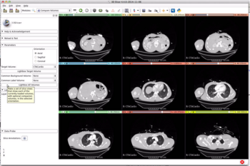
New Slice View Annotations is now available in DataProbe module. This new feature includes information about volume nodes of different layers of slice view in the form of corner text annotations (including DICOM information if available), color scalar bar and an interactive scaling ruler.
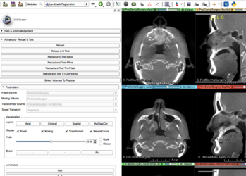
New LandmarkRegistration module providing interactive registration and inspection.
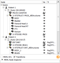
New Subject hierarchy module organizes and handles loaded data, providing processing and analysis features through plugins.
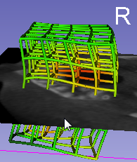
Improved Transforms module with support for non-linear transforms, visualization of transforms in 2D and 3D, detailed transform properties view - click here for demo video.
 New ExtensionsWizard tool deprecating the ModuleWizard.
- This new tool separates the concepts of extensions and modules, and allows creating an extension containing several modules, as well as adding modules to an existing extension.
- It allows to publish an extension source code to github.
- It provides an easy way to create a pull request on the extension index.
New Compare Volumes supports an overview of one or more volumes in the scene.
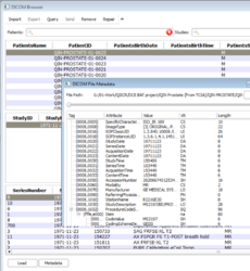
DICOM module user interface is improved for a better user experience. DICOM meta-data (header) viewer is now available and auto-examine in the DICOM browser makes it easier to work with plugins.

All application error and warning messages are saved to file. Logged messages for the current and recent sessions are available from the new error report box (menu: Help / Report a bug).
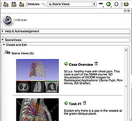
Scene Views module GUI updated with better layout.
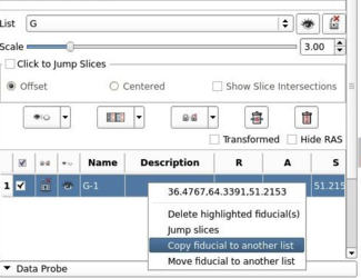
Markups module fiducial right click menu expanded to include copy and showing RAS coordinates. Show Slice Intersections check box added.
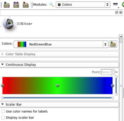
Colors module expanded to support continuous color maps, added an option to use the color names as labels on the scalar bar.


DICOM
- Improved DICOM support and user experience
- Added a way to change the table densities in three levels: compact, cozy and comfortable.
- Re-arranged the patient, study and series search box so that the search box will be on the top of tables for both horizontal and vertical cases.
- Resize tables based on contents.
- Allow users to acknowledge all DICOM loading errors with a single click
- Add DICOM meta-data (header) viewer
- Auto-examine in dicom browser added by providing two modes: Advanced and Non-advanced mode.
- Added repair tool for DICOM database
Transforms
- Improved integration of non rigid deformations, including grid (displacement field) and bspline transforms
- Interactive application of non rigid deformations to volume slices, models, markups
- Visualization of any transforms as glyphs, grid, or contours in 2D slice and 3D views - click here for demo video
- Computing and applying inverse transforms, compositing any number of transforms
- Real-time update: if the transform (or any visualization parameter) is changed then the visualization is updated immediately (interactive visualization while editing the transform)
- Detailed transform information display (type of transform, basic properties, displacement at current position)
- Loading/saving of oriented bspline transforms with or without additive bulk component
- Loading/saving of oriented grid transforms
- Loading/saving of transforms in h5 file format
Colors
- Improved Colors module user interface
- Added support to display continuous color scales
- Added an option to use color names as labels on the scalar bar
Markups
- Improved Markups module user interface
- Add slice intersections toggle
- Add right click option to copy markups list
- Add coordinates to right click menu
Editor
- Add a Sphere option to the PaintEffect to make it quicker to segment large anatomical regions that are somewhat spherical.
- Paint using pixel mode if brush size is too small
- Button effect are checked/unchecked based on the 'effect' property stored in the parameter node
Extensions Manager
- Add search box
- Add 'More' link
- Load extension icon from disk
- Implement downloading of extension icons
- Install extension dependencies
- Add mechanism to check for extensions updates
Slicer Extensions
- New and Improved Extensions
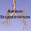
Airway Segmentation to segment the airway from chest CT images. UPDATED
CardiacAgastonMeasures to auto-segment the coronary calcium in Cardiac CT scans and then calculate the Agatston score and label statistics NEW

Cardiac MRI Toolkit to analyze cardiac LGE-MRI images. UPDATED

CarreraSliceInteractiveSegmenter to interactively segment in 3D UPDATED

CBC 3D (CRTC's BCC & Compression) I2M (Image-To-Mesh) Conversion for Image Guided Therapy. The extension encapsulates two CLI modules: (1) Body Centric Cubic (BCC) Mesh Generation. (2) Mesh Compression (MC). NEW
Change Tracker for quantification of the subtle changes in pathology. UPDATED
CleaverExtension extension bundles the Cleaver meshing tool as a CLI module NEW

Dice Computation to calculate the Dice Similarity Coefficient (DSC) between multiple label map images. UPDATED
CMFreg is a set of tools package for cranio-maxillofacial registration UPDATED
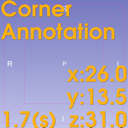
CornerAnnotation is a module to display annotations, time count, node elements on each panels on the Slicer user interface UPDATED

CurveMaker is a module to generate a curve based on a list of fiducial points. NEW
DTI Prep. UPDATED
DTI Process. UPDATED

FastGrowCut is an editor effect providing a fast implementation of the GrowCut method that supports multi-label segmentations UPDATED

FinslerTractography implements the Finsler tractography method with HARDI data as described by J. Melonakos et al., Finsler Active Contours, IEEE Trans PAMI, 30:412-423, 2008. NEW

GelDosimetryAnalysis is a Slicelet covering the gel dosimetry analysis workflow used in commissioning new radiation techniques. UPDATED

GyroGuide determines the trajectory angle and depth of puncture path, and transmits the calculated information to the probe for real time navigation. NEW

IASEM to segmentation and process of IASEM Electron Microscopy images. UPDATED

iGyne is an open source software for MR-Guided Interstitial Gynecologic Brachytherapy. NEW
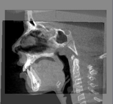
IntensitySegmenter is a simple tool that segments an image according to intensity value. It is mainly used to segment CT scans using the Hounsfield scale but the ranges of intensities and their corresponding labels can be specified in an input text file. UPDATED

LightWeightRobotIGT to manage communication between 3D Slicer and LightWeight robot. NEW
Longitudinal PET/CT to provide a user friendly Slicer interface for quantification of DICOM PET/CT image data.{{updated}

MABMIS: Multi-Atlas Based Group Segmentation NEW

Matlab Bridge to allow running Matlab functions directly in 3D Slicer. UPDATED
MultidimData: A set of modules for generic multidimensional data management in Slicer (0.2.1) NEW

NeedleFinder: NeedleFinder: fast interactive needle detection. It provides interactive tools to segment needles in MR/CT images. It has been mostly tested on MRI from gynelogical brachytherapy cases. NEW
PBNRR: This extension encapsulates a CLI module for the Physics-Based Non-Rigid Registration (PBNRR) method. The PBNRR compensates for the brain shift during the Image-Guided Neurosurgery (IGNS). NEW
PercutaneousApproachAnalysis: The Percutaneous Approach Analysis is used to calculate and visualize the accessibility of liver tumor with a percutaneous approach. NEW

PerkTutor for training in image-guided needle interventions. UPDATED

PETDICOM: The PET DICOM Extension provides tools to import PET Standardized Uptake Value images from DICOM into Slicer. NEW
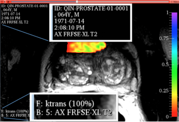
Pk Modeling to calculate quantitative parameters from Dynamic Contrast Enhanced DCE-MRI images. UPDATED
Port Placement to assists in the planning of surgical port placement in laparoscopic procedures. UPDATED

PyDevRemoteDebug: This extension allows remote visual debugging of Python scripts using PyDev (http://pydev.org/) NEW

Reporting to create image annotations/markup that are stored in a structured form. UPDATED

ResectionPlanner: Modules for surgical resection planning. NEW
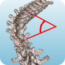
Scoliosis: Extensions pertaining to scoliosis analysis UPDATED

ShapePopulationViewer: Visualize and interact with multiple surfaces at the same time to easily compare them UPDATED

http://slicer.vmtk.org/ SlicerExtension-VMTK]: The Vascular Modeling Toolkit as a 3D Slicer4 extension. NEW
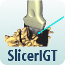
SlicerIGT to use all the advanced features of 3D Slicer for real-time navigation. UPDATED

SlicerRT is a tool for powerful radiotherapy research. UPDATED
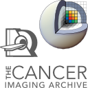
TCIABrowser: A Module to connect to TCIA archive, browse the collections, patients and studies and download DICOM files to 3D Slicer. NEW
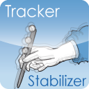
Tracker Stabilizer to output a filtered transform node based on an tracker input (transform node). UPDATED

UKF Tractography a framework which uses an unscented Kalman filter for performing tractography. UPDATED

VolumeClip: Clip volumes with surface models and ROI boxes NEW
Window/Level Effect to adjust window/level for volumes using mouse and/or region of interest. UPDATED
XNAT Slicer Secure GUI-based IO with any XNAT server. UPDATED


Other Improvements, Additions & Documentation
Optimization
- Improve Rendering pipeline performance optimizing observations management
- Reduce memory footprint (Fix memory leaks)
- Reduce installer size
- Faster image stack (png, jpg, bmp, tiff...) loading
Rendering / Visualization
- Add Multi-sampling option
CLI / SlicerExecutionModel
- Improved CLI AutoRun ensuring slice view is not reset when inputs are updated.
- In developer mode do not remove CLI input and output files
- Reduce the chance of crashes when CLI returns result images
Python scripting
- Add support for real Qt resources in Python. See r23290 for details.
- Improve VTK event support adding a way to specify the CallData type. See here for details.
- Bundle pydicom python module in Slicer package
- Add helper methods to
ScriptedLoadableModuleLogicfor managing parameter nodes
- SelfTest: Add
clickAndDrag()method to the scripted module. It allows to send synthetic mouse events to the specified widget (qMRMLSliceWidget or qMRMLThreeDView)
- Improve
slicer.utilpython module.- Add
modulePath(moduleName)method - Add
resetThreeDViews/resetSliceViews - Add
VTKObservationMixin - Add
getFirstNodeByClassAndName() - Add
NodeModifycontext manager: It allows to easily disable modified event associated with a node, and automatically re-enable them and invoking them if it applies.
- Add
- Markups: Add an event PointEndInteractionEvent for the end of a fiducial interaction
- TODO: Add reference to VTKv6 blog posts
Other
- Help / Report a bug: Application error/warning/debug log messages are now saved to file. Added option to copy/paste log file contents of recent Slicer sessions to bug reports.
For Developers
Under the hood
- VolumeLogic:
- Add method ResampleVolumeToReferenceVolume
- Add method CloneVolumeWithoutImageData
- Add support for user-defined stereo-viewing options
- Build-system
- Improve support for Visual Studio 2013
- Refactor management of external project launcher settings. See r23724
- Add option
Slicer_ITKV3_COMPATIBILITY. This option enabled by default will allow (if disabled) to build Slicer with ITKv3 compatibility later disabled andITK_USE_64BITS_IDSenabled.
- SlicerExecutionModel: Add ParameterSerializer support.
- Improved Toolkits

Moved from CTKAppLauncher v0.1.11 to v0.1.14 (43 commits)

Moved from ITK v4.4.1 to v4.6.0 (1089 commits)
Moved from OpenIGTLink 66e272d to 849b434 (53 commits)

Moved from Qt 4.7.4 to Qt 4.8.6

Moved from VTK v5.10.1 to VTK v6.2.0 (5490 commits)
Looking at the Code Changes
From a git checkout you can easily see the all the commits since the time of the 4.4.0 release:
git log v4.3.0..HEAD
To see a summary of your own commits, you could use something like:
git log v4.3.0..HEAD --oneline --author=pieper
see the git log man page for more options.
Commit stats and full changelog