Documentation/4.8/Modules/BrainStructuresSegmenter
|
For the latest Slicer documentation, visit the read-the-docs. |
Introduction and Acknowledgements
|
Extension: BrainTissuesExtension | |||||||
|
Module Description
This module offers a general brain segmentation pipeline, calling different algorithms depending on the tissue type that the user wants to segment. The list below illustrates the segmentation methods available at moment:
- Basic Brain Tissues: A general brain tissues segmentation focused on the white matter, gray matter and CSF brain tissue segmentation.
- Brain Logistic Classification (BLS): A recent brain segmentation method focused on global tissue segmentation, usually CSF, gray and white matter delineations. general brain tissues segmentation focused on the white matter, gray matter and CSF brain tissues segmentation.[1]
NOTE: Each segmentation method listed are related to the segmentation part of the image processing pipeline controlled by this module, i.e. the Brain Structure Segmenter module apply a set of image processing algorithms in which, in the Segmentation part, uses the above methods. The user still has the flexibility to choose what pre and post processing that should be applied in the general image processing pipeline.
The diagram below illustrates the image processing pipeline used in this module

Use Cases
- Use Case 1: Tissue classification
- There are several image quantitative approaches that use only a certain tissue type (for instance, cortical thickness) in which a previous brain segmentation could be needed.
- A simple brain tissue mask could be obtained from this module (WM, GM and CSF are available at moment).
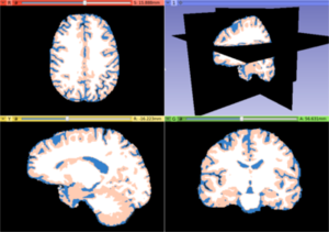
White matter, gray matter and CSF tissues segmented from the previous MRI image
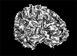
A white matter mask 3D reconstruction
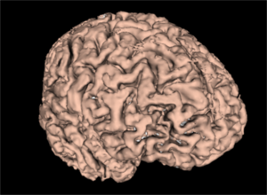
A gray matter mask 3D reconstruction


Panels and their use

IO:
- Input Volume
- Pick the input to the algorithm. This should be an MRI strutural images with a type listed in the Image Modality option
- Image Modality
- MRI strutural image inserted as a input volume
- Is brain extracted?
- Is the input data already brain extracted? If not, the ROBEX brain extraction method is used
- Output Volume
- Pick the output to the algorithm (a label image)
Tissue Segmentation Parameters:
- Separate one tissue?
- Select one tissue type desired to be passed as the output. If checked, the tissue type in Tissue Type option is used
- Tissue Type
- Tissue type that will be resulted from the brain segmentation
Noise Attenuation Parameters:
- Condutance
- The conductance regulates the diffusion intensity in the neighbourhood area. Choose a higher conductance if the input image has strong noise seem in the whole image space
- Number Of Iterations
- The number of iterations regulates the numerical simulation of the anomalous process over the image. This parameters is also related with the de-noising intensity, however it is more sensible to the noise intensity. Choose the higher number of iterations if the image presents high intensity noise which is not well treated by the conductance parameter
- Q Value
- The anomalous parameter (or q value) is the generalization parameters responsible to give the anomalous process approach on the diffusion equation
Label Refinement Parameters
- Gaussian Sigma
- Label smoothing by a gaussian distribution with variance sigma. The units here is given in mm
Similar Modules
References
N/A
Information for Developers
| Section under construction. |
- ↑ BLS paper