Home < Documentation < 4.4 < Modules < T1 Mapping
For the latest Slicer documentation, visit the 4.10 page.
|
Introduction and Acknowledgements
Extension: T1_Mapping
Acknowledgments:
This work is part of the National Alliance for Medical Image Computing (NA-MIC), funded by the National Institutes of Health through the NIH Roadmap for Medical Research, and by National Cancer Institute as part of the Quantitative Imaging Network initiative (U01CA154601) and QIICR (U24CA180918).
Implementation of the T1 Mapping was contributed by Xiao Da from MGH.
Author: Xiao Da (MGH), Artem Mamonov (MGH), Jayashree Kalpathy-Cramer (MGH) and Andriy Fedorov (BWH)
Contact: Xiao Da, <email>XDA@mgh.harvard.edu</email>
|
| National Alliance for Medical Image Computing (NA-MIC)
|
| Quantitative Image Informatics for Cancer Research
|
| Martinos Center for Biomedical Imaging
|
|
|
Module Description
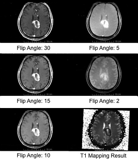
T1 mapping estimates effective tissue parameter maps (T1) from multi-spectral FLASH MRI scans with different flip angles.
Use Cases
- Take multi-spectral FLASH images with an arbitrary number of flip angles as input, and estimate the T1 values of the data for each voxel.
- Read repetition time(TR), echo time(TE) and flip angles from the Dicom header directly.
- Prostate, brain, head & neck, cervix, breast and etc.
Sample Results of T1 Mapping
|
Tutorials
Panels and their use
|
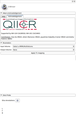
- Parameters:
- Input Volume: Multi-spectral FLASH MRI on multi-volume dicom format
- Output Volume: T1 mapping result calculated at each voxel
|
Similar Modules
References
Information for Developers
Source code: https://github.com/stevedaxiao/T1_Mapping