Difference between revisions of "EMSegmenter-Tasks:CT-Hand-Bone"
Belhachemi (talk | contribs) |
Belhachemi (talk | contribs) (→Atlas) |
||
| (17 intermediate revisions by 2 users not shown) | |||
| Line 2: | Line 2: | ||
=Description= | =Description= | ||
| − | Single channel automatic segmentation of CT hand scans into the finger bones. The task can be applied to right and and left hand scans. | + | Single channel automatic segmentation of CT hand scans into the finger bones based on the publication (Ramme et al). The task can be applied to right and and left hand scans. Subjects should be scanned with a similar protocol as atlas, which is .... |
| − | |||
| − | |||
| − | |||
| − | |||
| − | |||
| − | |||
| − | |||
| − | |||
| − | |||
| − | |||
| − | |||
| − | |||
| − | |||
| − | |||
| − | |||
| − | |||
| − | |||
| − | |||
| − | |||
| − | |||
| − | |||
| − | |||
| − | |||
| − | |||
| − | |||
| − | |||
| + | The pipeline consist of the following steps: | ||
| + | * Step 1: Flip/Mirror the atlas (based on right hand scans) if scan of the subject is of the left hand. | ||
| + | * Step 2: Create Binary MAP of the hand for the atlas template and CT scan. For each scan: | ||
| + | ** Generate binary map of the scan by thresholding the scan above 150 | ||
| + | ** Fill holes in binary map by | ||
| + | *** Blurring via itk::DiscreteGaussianImageFilter (parameter Variance=1.5, MaximumKernelWidth 5) | ||
| + | *** Binarize map via itk::BinaryThresholdImageFilter (Set everything between 0 and 30 to 0, otherwise 255) | ||
| + | ** Remove noise from the binary map by extracting largest component | ||
| + | <!--itk::BinaryThresholdImageFilter to extract label 255. keep everything between 1 and 255 (1-254 are not used) and set it to one --> | ||
| + | *** apply itk::ConnectedComponentImageFilter to label the objects in the binary image. Each distinct object is assigned a unique label. | ||
| + | *** Use itk::RelabelComponentImageFilter to sort the labels based on the size of the object: the largest object will have label #1, the second largest will have label #2, etc. | ||
| + | *** Use itk::BinaryThresholdImageFilter to extract label 1. This is the largest object in the data set. | ||
<!-- | <!-- | ||
Island Removal and Fill: | Island Removal and Fill: | ||
| Line 38: | Line 24: | ||
Binary Threshold | Binary Threshold | ||
--> | --> | ||
| − | + | * Step 3: Register the binarized atlas template to the binarized CT scan via [[Modules:BRAINSFit| BRAINSFit]] (Johnson et al 2007) | |
| − | + | ** Step A: Use BRAINSFit to perform a affine registration. Register the atlas template linear to the subject scan and save the linear transformation. (BRAINSFit Rigid,Affine) | |
| − | + | ** Step B: Use BRAINSDemonWarp to perform a non-linear registration. Register the atlas template non-linear to the subject scan. Use the linear transformation as initialization. (BRAINSDemonWarp) | |
| − | * Step | + | ** Step C: Use BRAINSResample together with the --deformationVolume option to resample the atlas files shown in the figure below. |
| − | ** Step | + | * Step 4: Compute the intensity distributions for each structure <BR> |
| − | |||
| − | |||
| − | |||
| − | ** Step | ||
| − | |||
| − | |||
| − | |||
| − | ** Step | ||
| − | * Step | ||
Compute intensity distribution (mean and variance) for each label by automatically sampling from the MR scan. The sampling for a specific label is constrained to the region that consists of voxels with high probability (top 95%) of being assigned to the label according to the aligned atlas. | Compute intensity distribution (mean and variance) for each label by automatically sampling from the MR scan. The sampling for a specific label is constrained to the region that consists of voxels with high probability (top 95%) of being assigned to the label according to the aligned atlas. | ||
| − | * Step | + | * Step 5: Generate a spatial atlas for tissue + remaining bones and air |
| + | * Step 6: Automatically segment the CT scan into the structures of interest using [[Modules:EMSegmenter-3.6|EM Algorithm]] (Pohl et al 2007) | ||
=Anatomical Tree= | =Anatomical Tree= | ||
| Line 85: | Line 63: | ||
=Atlas= | =Atlas= | ||
| + | |||
| + | The task is taking advantage of fuzzy atlas data for the 12 finger bones. | ||
| + | |||
| + | <gallery perrow=1: widths=630px : heights=280px> | ||
| + | Image:EMS_Atlas_IndexFinger2.png|fuzzy atlas for each finger bone <br> In this picture from left to right: index finger / digitus secundus (II): 'Distal (II)' 'Medial (II)' 'Proximal (II)' | ||
| + | </gallery> | ||
=Result= | =Result= | ||
| − | <gallery perrow=1: widths=630px : heights=630px> | + | <gallery perrow=1: widths=630px : heights=280px> |
| − | Image:EMS_Result_3DReconstruction_CT-Hand-Bone.png | + | Image:EMS Result Hand subject2.png|label map produced by the EMSegmenter |
| + | </gallery> | ||
| + | |||
| + | <!-- | ||
| + | Image:CTHandBone_BeforeAfter.png|3D reconstruction of the label map | ||
| + | --> | ||
| + | |||
| + | <gallery perrow=1: widths=630px : heights=600px> | ||
| + | Image:EMS_Result_3DReconstruction_CT-Hand-Bone.png|3D reconstruction of the label map overlayed with the opaque hand tissue | ||
</gallery> | </gallery> | ||
| Line 103: | Line 95: | ||
* Johnson H.J., Harris G., Williams K. [http://hdl.handle.net/1926/1291 BRAINSFit: Mutual Information Registrations of Whole-Brain 3D Images, Using the Insight Toolkit], The Insight Journal, July 2007 | * Johnson H.J., Harris G., Williams K. [http://hdl.handle.net/1926/1291 BRAINSFit: Mutual Information Registrations of Whole-Brain 3D Images, Using the Insight Toolkit], The Insight Journal, July 2007 | ||
* T. Vercauteren, X. Pennec, A. Perchant, N. Ayache. Symmetric Log-Domain Diffeomorphic Registration: A Demons-based Approach. MICCAI 2008 | * T. Vercauteren, X. Pennec, A. Perchant, N. Ayache. Symmetric Log-Domain Diffeomorphic Registration: A Demons-based Approach. MICCAI 2008 | ||
| + | * Ramme AJ, Devries N, Kallemyn NA, Magnotta VA, Grosland NM. [http://www.springerlink.com/content/h972u63630311g78/ Semi-automated Phalanx Bone Segmentation Using the Expectation Maximization Algorithm]. J Digit Imaging. 2008. | ||
Latest revision as of 16:11, 5 May 2011
Home < EMSegmenter-Tasks:CT-Hand-BoneReturn to EMSegmenter Task Overview Page
Contents
Description
Single channel automatic segmentation of CT hand scans into the finger bones based on the publication (Ramme et al). The task can be applied to right and and left hand scans. Subjects should be scanned with a similar protocol as atlas, which is ....
The pipeline consist of the following steps:
- Step 1: Flip/Mirror the atlas (based on right hand scans) if scan of the subject is of the left hand.
- Step 2: Create Binary MAP of the hand for the atlas template and CT scan. For each scan:
- Generate binary map of the scan by thresholding the scan above 150
- Fill holes in binary map by
- Blurring via itk::DiscreteGaussianImageFilter (parameter Variance=1.5, MaximumKernelWidth 5)
- Binarize map via itk::BinaryThresholdImageFilter (Set everything between 0 and 30 to 0, otherwise 255)
- Remove noise from the binary map by extracting largest component
- apply itk::ConnectedComponentImageFilter to label the objects in the binary image. Each distinct object is assigned a unique label.
- Use itk::RelabelComponentImageFilter to sort the labels based on the size of the object: the largest object will have label #1, the second largest will have label #2, etc.
- Use itk::BinaryThresholdImageFilter to extract label 1. This is the largest object in the data set.
- Step 3: Register the binarized atlas template to the binarized CT scan via BRAINSFit (Johnson et al 2007)
- Step A: Use BRAINSFit to perform a affine registration. Register the atlas template linear to the subject scan and save the linear transformation. (BRAINSFit Rigid,Affine)
- Step B: Use BRAINSDemonWarp to perform a non-linear registration. Register the atlas template non-linear to the subject scan. Use the linear transformation as initialization. (BRAINSDemonWarp)
- Step C: Use BRAINSResample together with the --deformationVolume option to resample the atlas files shown in the figure below.
- Step 4: Compute the intensity distributions for each structure
Compute intensity distribution (mean and variance) for each label by automatically sampling from the MR scan. The sampling for a specific label is constrained to the region that consists of voxels with high probability (top 95%) of being assigned to the label according to the aligned atlas.
- Step 5: Generate a spatial atlas for tissue + remaining bones and air
- Step 6: Automatically segment the CT scan into the structures of interest using EM Algorithm (Pohl et al 2007)
Anatomical Tree
The anatomical tree represents the structures to be segmented. Node labels displayed below contain a human readable structure name and in parentheses the internally used structure name.
- Hand
- Air
- Tissue
- Index finger / digitus secundus (II)
- Proximal (II)
- Medial (II)
- Distal (II)
- Middle finger / digitus medius (III)
- Proximal (III)
- Medial (III)
- Distal (III)
- Ring finger / digitus annularis (IV)
- Proximal (IV)
- Medial (IV)
- Distal (IV)
- Little finger / digitus minimus (V)
- Proximal (V)
- Medial (V)
- Distal (V)
Atlas
The task is taking advantage of fuzzy atlas data for the 12 finger bones.
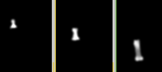
fuzzy atlas for each finger bone
In this picture from left to right: index finger / digitus secundus (II): 'Distal (II)' 'Medial (II)' 'Proximal (II)'

Result
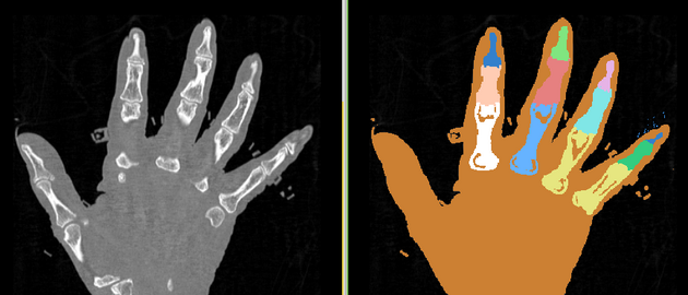
label map produced by the EMSegmenter

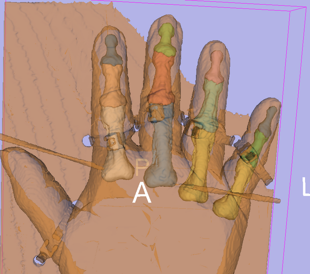
3D reconstruction of the label map overlayed with the opaque hand tissue

Collaborators
Vincent Magnotta (University of Iowa)
Acknowledgment
The construction of the pipeline was supported by funding from NIH NCRR 2P41RR013218 Supplement.
Citations
- Pohl K, Bouix S, Nakamura M, Rohlfing T, McCarley R, Kikinis R, Grimson W, Shenton M, Wells W. A Hierarchical Algorithm for MR Brain Image Parcellation. IEEE Transactions on Medical Imaging. 2007 Sept;26(9):1201-1212.
- S. Warfield, J. Rexilius, P. Huppi, T. Inder, E. Miller, W. Wells, G. Zientara, F. Jolesz, and R. Kikinis, “A binary entropy measure to assess nonrigid registration algorithms,” in MICCAI, LNCS, pp. 266–274, Springer, October 2001.
- Johnson H.J., Harris G., Williams K. BRAINSFit: Mutual Information Registrations of Whole-Brain 3D Images, Using the Insight Toolkit, The Insight Journal, July 2007
- T. Vercauteren, X. Pennec, A. Perchant, N. Ayache. Symmetric Log-Domain Diffeomorphic Registration: A Demons-based Approach. MICCAI 2008
- Ramme AJ, Devries N, Kallemyn NA, Magnotta VA, Grosland NM. Semi-automated Phalanx Bone Segmentation Using the Expectation Maximization Algorithm. J Digit Imaging. 2008.