File list
From Slicer Wiki
This special page shows all uploaded files.
| Date | Name | Thumbnail | Size | User | Description | Versions |
|---|---|---|---|---|---|---|
| 19:14, 15 January 2013 | Pujol-AcademicRadiology2008-fig1.jpg (file) |  |
533 KB | Marianna | 1 | |
| 19:07, 15 January 2013 | Maier-MICCAI2011-fig4.jpg (file) |  |
183 KB | Marianna | 1 | |
| 17:44, 15 January 2013 | Irimia-JNeurotrauma2011-fig2.jpg (file) |  |
211 KB | Marianna | 1 | |
| 17:33, 15 January 2013 | Vikal-ComputMedImagingGraph2009-fig5.jpg (file) |  |
132 KB | Marianna | 1 | |
| 17:33, 15 January 2013 | Vikal-ComputMedImagingGraph2009.pdf (file) |  |
7.27 MB | Marianna | 1 | |
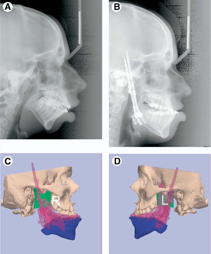
| 17:28, 15 January 2013 | Kaban-JOralMaxillofacSurg2009-fig7.jpg (file) |  |
266 KB | Marianna | 1 | |
| 17:27, 15 January 2013 | Lindig-HistochemCellBiol2009-fig6.pdf (file) |  |
18.84 MB | Marianna | 1 | |
| 17:16, 15 January 2013 | Xia-IJMR2008-fig6.jpg (file) | 264 KB | Marianna | 1 | ||
| 17:16, 15 January 2013 | Xia-IntJMedRoboticsComputAssistSurg2008.pdf (file) |  |
3.46 MB | Marianna | 1 | |
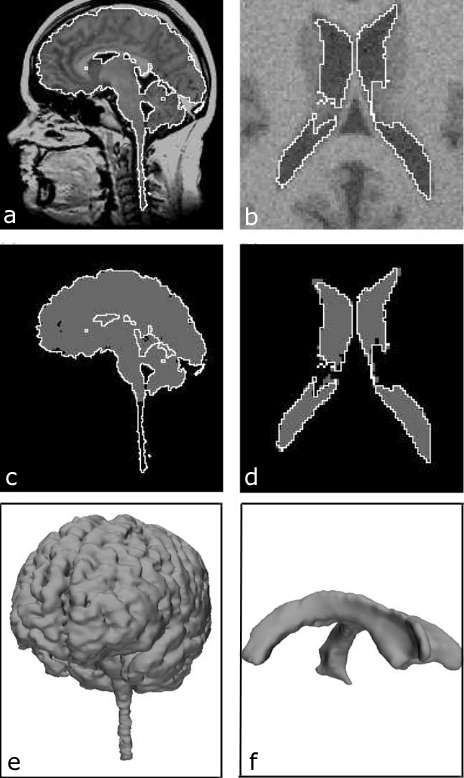
| 17:06, 15 January 2013 | Nakajima-Neuroimage2007-fig7.jpg (file) |  |
47 KB | Marianna | 1 | |
| 17:06, 15 January 2013 | Nakajima-Neuroimage2007.pdf (file) |  |
7.54 MB | Marianna | 1 | |
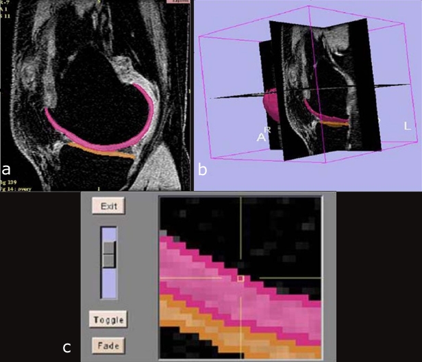
| 17:01, 15 January 2013 | Brem-SkeletalRadiol2007-fig1.jpg (file) |  |
151 KB | Marianna | 1 | |
| 17:01, 15 January 2013 | Brem-SkeletalRadiology2007.pdf (file) | 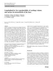 |
191 KB | Marianna | 1 | |
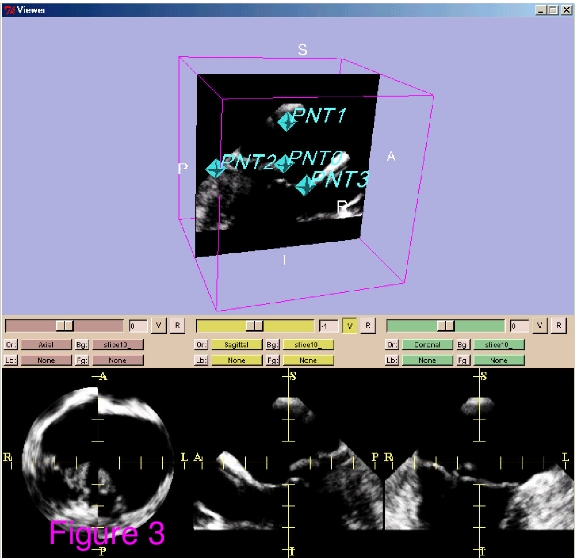
| 16:50, 15 January 2013 | VerheyBioMedEngOnline2006-fig3.jpg (file) |  |
126 KB | Marianna | 1 | |
| 16:49, 15 January 2013 | VerheyBioMedEngOnline2006.pdf (file) | 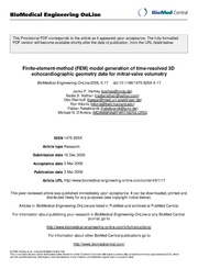 |
1.14 MB | Marianna | 1 | |
| 16:34, 15 January 2013 | Pichon-MedIA2004-fig2.jpg (file) |  |
145 KB | Marianna | 1 | |
| 16:29, 15 January 2013 | McConnell-Neurosurgery2004-fig1.jpg (file) |  |
430 KB | Marianna | 1 | |
| 16:23, 15 January 2013 | Nabavi-ActaNeurochir2002-fig1.jpg (file) |  |
444 KB | Marianna | 1 | |
| 16:23, 15 January 2013 | Nabavi-ActaNeurochir2002.pdf (file) | 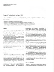 |
4.21 MB | Marianna | 1 | |
| 16:16, 15 January 2013 | Rodt-Neuroradiology2002-fig5.jpg (file) |  |
144 KB | Marianna | 1 | |
| 16:16, 15 January 2013 | Rodt-Neuroradiology2002.pdf (file) | 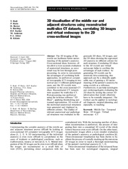 |
458 KB | Marianna | 1 | |
| 16:00, 15 January 2013 | Nabavi-Neurosurgery2001-fig3.jpg (file) |  |
190 KB | Marianna | 1 | |
| 15:59, 15 January 2013 | Nabavi-Neurosurgery2001.pdf (file) |  |
330 KB | Marianna | 1 | |
| 17:14, 11 January 2013 | Egger-pone.0051788-2012-fig1.png (file) |  |
397 KB | Marianna | 1 | |
| 16:52, 11 January 2013 | Rodt-ChildsNervSyst2007.pdf (file) |  |
310 KB | Marianna | 1 | |
| 16:52, 11 January 2013 | Rodt-ChildsNervSyst2007-fig3.png (file) |  |
243 KB | Marianna | 1 | |
| 15:28, 11 January 2013 | Rodrigues-AmJObstetGynecol2011.pdf (file) |  |
1.79 MB | Marianna | 1 | |
| 15:18, 11 January 2013 | Fedorov 3DSlicer MRI.pdf (file) |  |
3.62 MB | Marianna | 1 | |
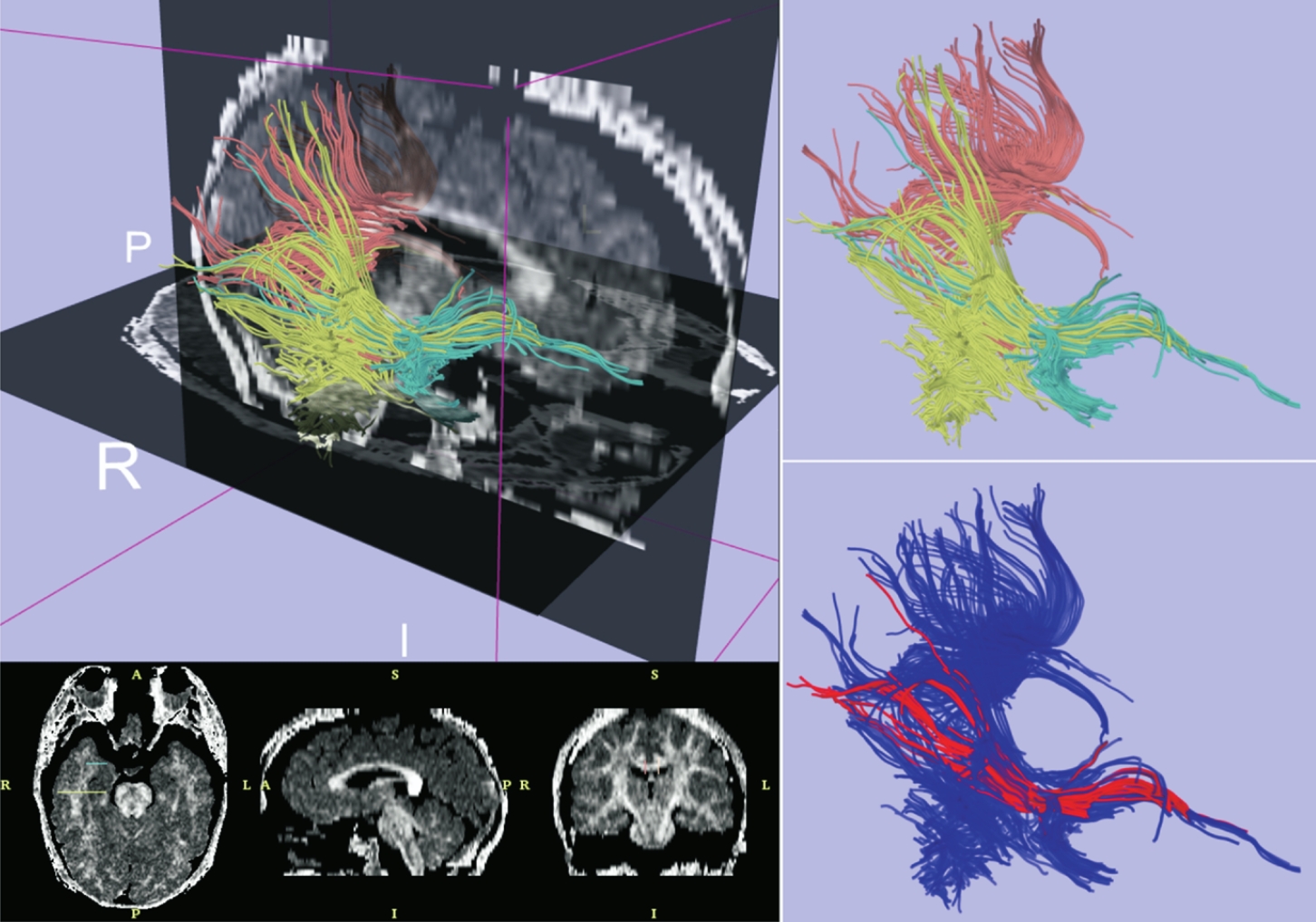
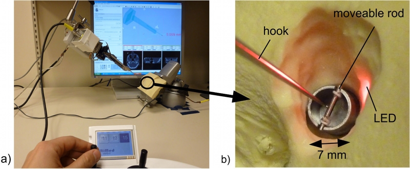
| 15:16, 11 January 2013 | Pinter-MedicalPhysics2012.pdf (file) |  |
1.56 MB | Marianna | 1 | |
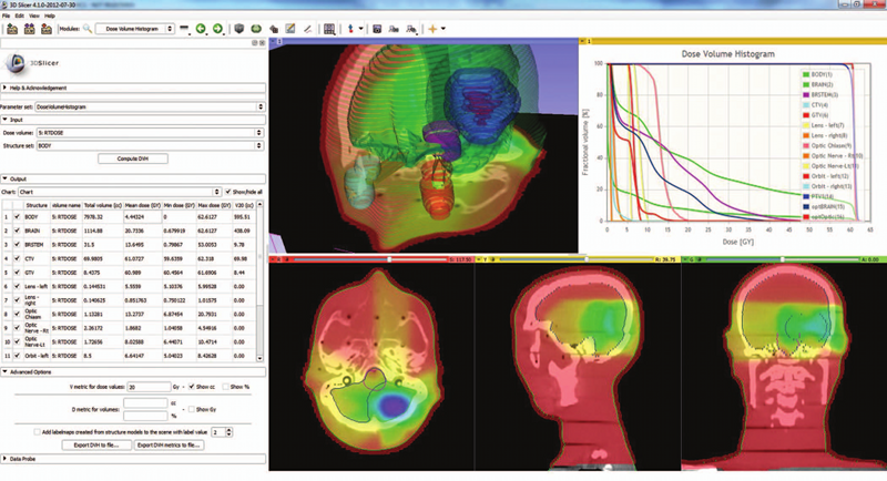
| 15:14, 11 January 2013 | Pinter-MedicalPhysics2012-fig2.png (file) |  |
464 KB | Marianna | 1 | |
| 22:30, 10 January 2013 | Huang-BMCMedImaging2008.pdf (file) | 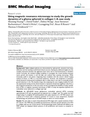 |
4.82 MB | Marianna | 1 | |
| 22:28, 10 January 2013 | Huang-BMC-MedImg2008-fig1.jpg (file) |  |
128 KB | Marianna | 1 | |
| 15:59, 9 January 2013 | LineMarkerRegistration Screenshot Confirmation.png (file) |  |
175 KB | Tokuda | 1 | |
| 15:58, 9 January 2013 | LineMarkerRegistration Screenshot ApplyTransform.png (file) | 74 KB | Tokuda | 1 | ||
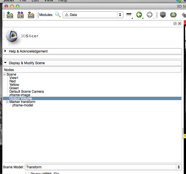
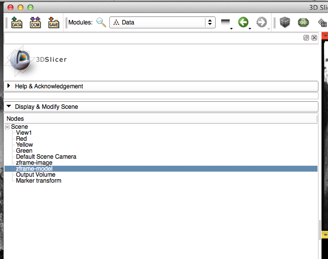
| 15:58, 9 January 2013 | LineMarkerRegistration Screenshot DataModule.png (file) |  |
66 KB | Tokuda | 1 | |
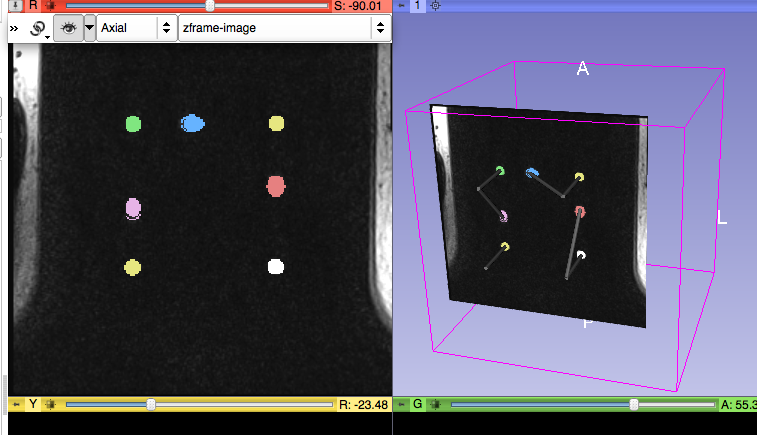
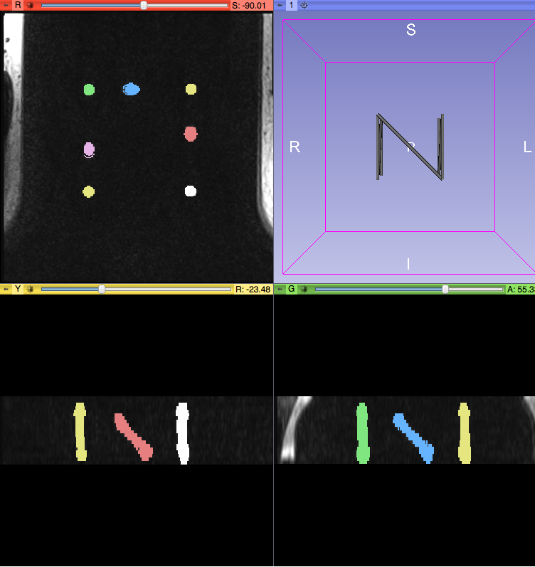
| 15:58, 9 January 2013 | LineMarkerRegistration Screenshot Segmented.png (file) |  |
184 KB | Tokuda | 1 | |
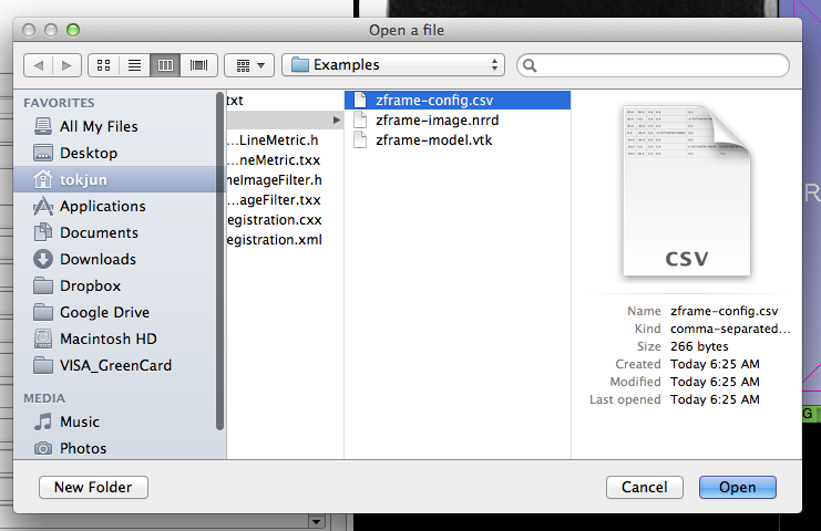
| 15:57, 9 January 2013 | LineMarkerRegistration Screenshot LoadCSV.png (file) |  |
114 KB | Tokuda | 1 | |
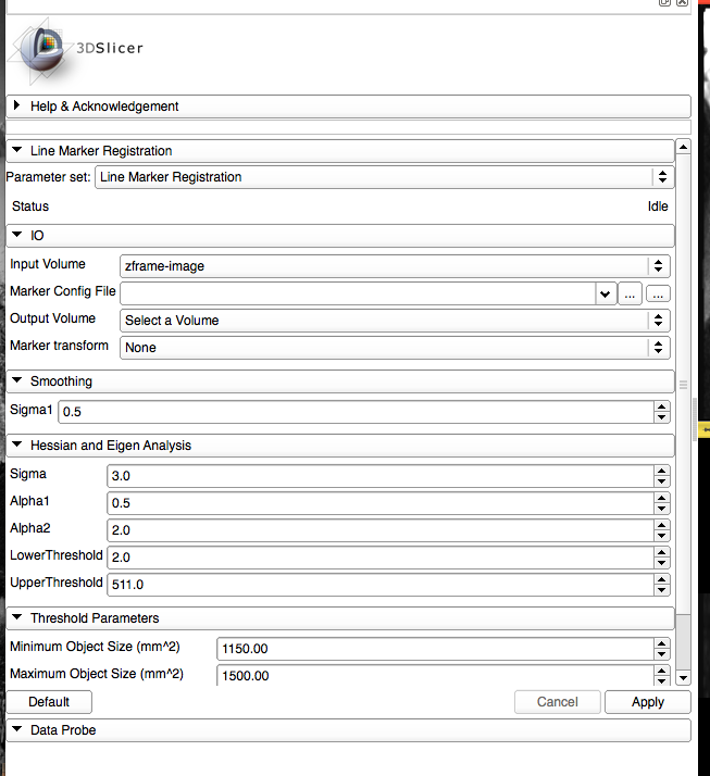
| 15:57, 9 January 2013 | LineMarkerRegistration Screenshot GUI.png (file) |  |
86 KB | Tokuda | 1 | |
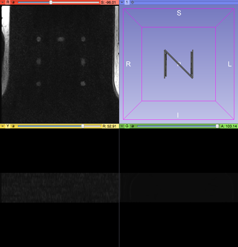
| 15:55, 9 January 2013 | LineMarkerRegistration Screenshot VisualizeData.png (file) |  |
168 KB | Tokuda | 1 | |
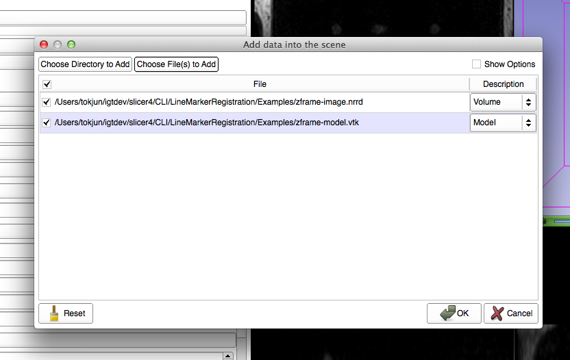
| 15:54, 9 January 2013 | LineMarkerRegistration Screenshot LoadData.png (file) |  |
88 KB | Tokuda | 1 | |
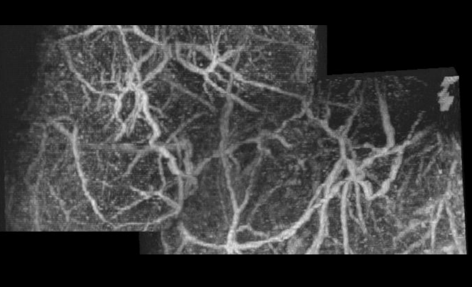
| 01:10, 9 January 2013 | Tumor final merge tubetk.png (file) |  |
307 KB | Crmullins | Using the TubeTK extension module "Merge (TubeTK)" we were able to successfully merge the tumor ultrasound images taken from the left and the right sides. | 1 |
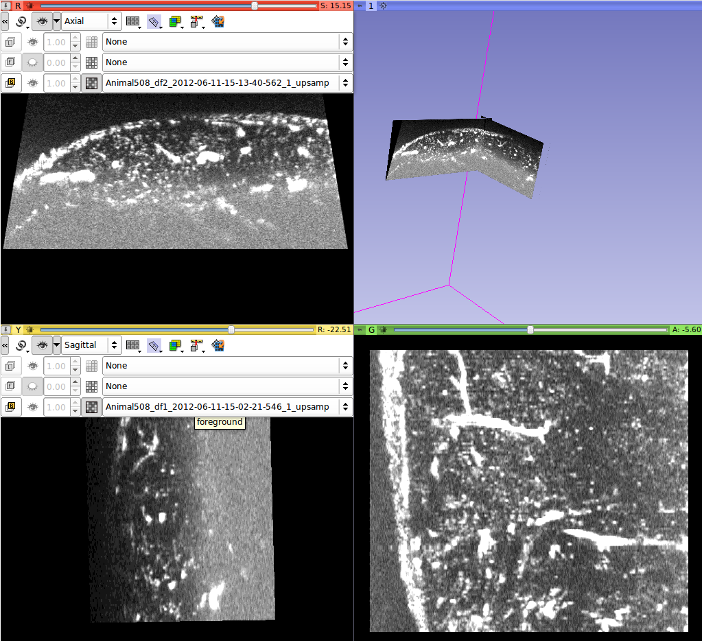
| 00:19, 9 January 2013 | Aligned tumor image-Axial Sagittal.png (file) |  |
254 KB | Crmullins | Screenshot of two tumor volumes (left and right) registered and transformed using Slicer's transforms module. Axial and sagittal shown to illustrate the lineup from front to back. | 1 |
| 00:15, 9 January 2013 | Aligned tumor image Axial-Axial.png (file) |  |
359 KB | Crmullins | A left and right tumor ultrasound registered and transformed using the Slicer transforms module. | 1 |
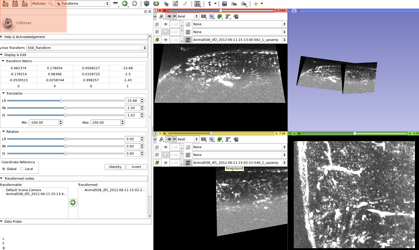
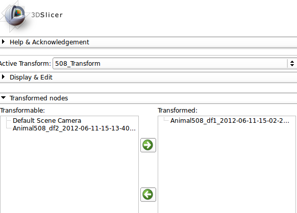
| 00:11, 9 January 2013 | Transforms module for tumor merges.png (file) | 26 KB | Crmullins | Screenshot of the transforms module for use in the UNC Prepalpable Tumor study. | 1 | |
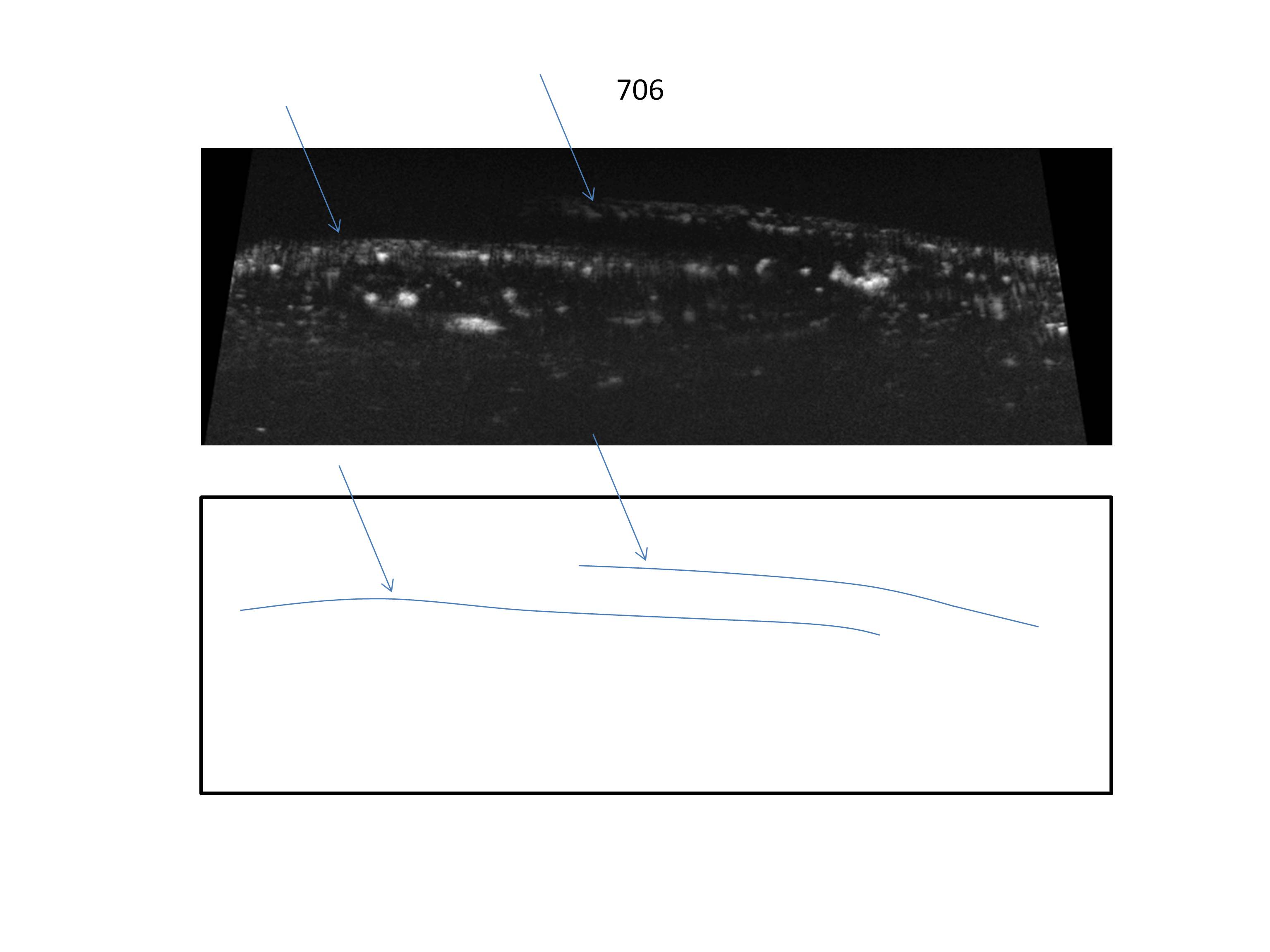
| 23:22, 8 January 2013 | 706 misregistration.jpg (file) |  |
224 KB | Crmullins | This is the result of using the tubeMerge module from the TubeTK extension to merge two ultrasound images which have not been properly registered. Notice the shadowing artifacts. | 1 |
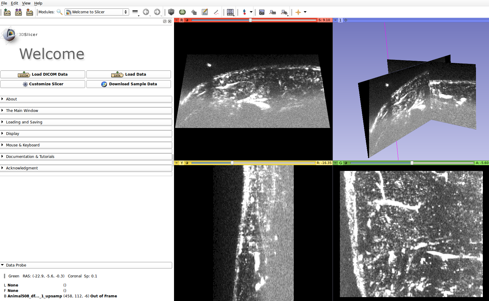
| 23:19, 8 January 2013 | Unaligned tumor image.png (file) |  |
383 KB | Crmullins | This is the result of loading two images to be registered simultaneously, before any transformation has been applied to the moving image. The next step is to use the Transforms module to apply a transform to the moving image. | 1 |
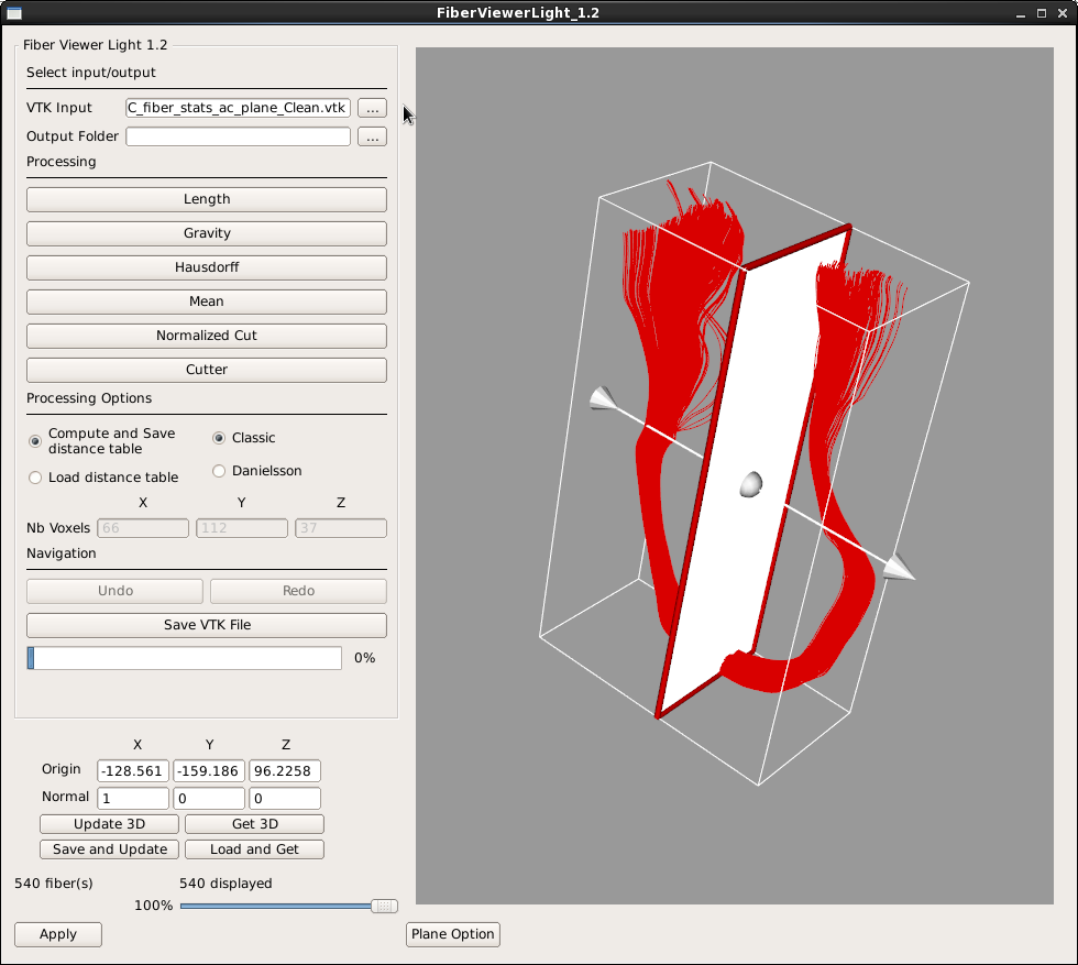
| 21:03, 2 January 2013 | FiberViewerLight1.2-ScreenShot.png (file) |  |
105 KB | Fbudin | 1 | |
| 20:16, 2 January 2013 | UNC-NIRAL.png (file) |  |
54 KB | Fbudin | logo | 1 |
| 19:50, 26 December 2012 | DICOM-architecture.png (file) |  |
65 KB | Pieper | 1 | |
| 22:41, 17 December 2012 | ProstateDx-01-0006-1.zip (file) | 4.43 MB | Spujol | 1 |