Difference between revisions of "Slicer3.4:Training"
From Slicer Wiki
| Line 79: | Line 79: | ||
{| border="1" cellpadding="5" | {| border="1" cellpadding="5" | ||
|- style="background:#849f66; color:#3b3f33; font-size:130%" align="center" | |- style="background:#849f66; color:#3b3f33; font-size:130%" align="center" | ||
| − | | style="width:8%" ;align="Center"| ''' | + | | style="width:8%" ;align="Center"| '''Driving Biological Project/Collaboration''' |
| style="width:40%" ;align="Center"| '''Tutorial''' | | style="width:40%" ;align="Center"| '''Tutorial''' | ||
| style="width:40%" ;align="Center"| '''Sample Data''' | | style="width:40%" ;align="Center"| '''Sample Data''' | ||
| style="width:12%" ;align="Center"| '''Image''' | | style="width:12%" ;align="Center"| '''Image''' | ||
|- | |- | ||
| − | | style="background:#849f66; color:#3b3f33; font-size:110%" align="Center"| <span id="1.1"></span> ''' | + | | style="background:#849f66; color:#3b3f33; font-size:110%" align="Center"| <span id="1.1"></span> '''Confocal Microscopy''' |
| style="background:#CCFF99; color:black" | '''[http://www.na-mic.org/Wiki/images/4/4d/Microscopy-Confocal-TrainingTutorial-2009JUNE.pdf Confocal Microscopy] ''' | | style="background:#CCFF99; color:black" | '''[http://www.na-mic.org/Wiki/images/4/4d/Microscopy-Confocal-TrainingTutorial-2009JUNE.pdf Confocal Microscopy] ''' | ||
| style="background:#CCFF99; color:black" | [http://www.ccdb.ucsd.edu/index.shtm Microscopy Confocal Dataset] | | style="background:#CCFF99; color:black" | [http://www.ccdb.ucsd.edu/index.shtm Microscopy Confocal Dataset] | ||
| style="background:#CCFF99; color:black" align="Center"| [[Image:MicroscopyTutorial.png| 250px]] | | style="background:#CCFF99; color:black" align="Center"| [[Image:MicroscopyTutorial.png| 250px]] | ||
|- | |- | ||
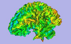
| − | | style="background:#849f66; color:#3b3f33; font-size:110%" align="Center"| <span id="1.1"></span> ''' | + | | style="background:#849f66; color:#3b3f33; font-size:110%" align="Center"| <span id="1.1"></span> '''Autism''' |
| style="background:#CCFF99; color:black" | '''[http://www.na-mic.org/Wiki/index.php/File:ARCTIC-Slicer3-Tutorial.pdf ARCTIC: Automatic Regional Cortical ThICkness V2.1] ''' | | style="background:#CCFF99; color:black" | '''[http://www.na-mic.org/Wiki/index.php/File:ARCTIC-Slicer3-Tutorial.pdf ARCTIC: Automatic Regional Cortical ThICkness V2.1] ''' | ||
| style="background:#CCFF99; color:black" | [http://www.nitrc.org/projects/arctic/ ARCTIC Data] | | style="background:#CCFF99; color:black" | [http://www.nitrc.org/projects/arctic/ ARCTIC Data] | ||
| style="background:#CCFF99; color:black" align="Center"| [[Image:Corticalthickness.png| 250px ]] | | style="background:#CCFF99; color:black" align="Center"| [[Image:Corticalthickness.png| 250px ]] | ||
|- | |- | ||
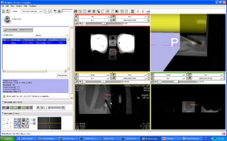
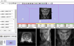
| − | | style="background:#849f66; color:#3b3f33; font-size:110%" align="Center"| <span id="1.1"></span> ''' | + | | style="background:#849f66; color:#3b3f33; font-size:110%" align="Center"| <span id="1.1"></span> '''Prostate''' |
| style="background:#CCFF99; color:black" | '''[http://www.na-mic.org/Wiki/images/0/06/DBP2JohnsHopkinsTransRectalProstateBiopsy_TutorialPres2009June.pdf Trans-rectal MR guided prostate biopsy] ''' | | style="background:#CCFF99; color:black" | '''[http://www.na-mic.org/Wiki/images/0/06/DBP2JohnsHopkinsTransRectalProstateBiopsy_TutorialPres2009June.pdf Trans-rectal MR guided prostate biopsy] ''' | ||
| style="background:#CCFF99; color:black" | [[Media:TransRectalProstateBiopsyTutorialDataset.zip| MR guided prostate biopsy Dataset]] | | style="background:#CCFF99; color:black" | [[Media:TransRectalProstateBiopsyTutorialDataset.zip| MR guided prostate biopsy Dataset]] | ||
| style="background:#CCFF99; color:black" align="Center"| [[Image:TransRectalBiopsy.png| 250px]] | | style="background:#CCFF99; color:black" align="Center"| [[Image:TransRectalBiopsy.png| 250px]] | ||
|- | |- | ||
| − | | style="background:#849f66; color:#3b3f33; font-size:110%" align="Center"| <span id="1.1"></span> ''' | + | | style="background:#849f66; color:#3b3f33; font-size:110%" align="Center"| <span id="1.1"></span> '''Schizophrenia''' |
| style="background:#CCFF99; color:black" | [[media:Stochastic_June09_3.ppt| '''Python Stochastic Tractography Module''']] | | style="background:#CCFF99; color:black" | [[media:Stochastic_June09_3.ppt| '''Python Stochastic Tractography Module''']] | ||
| style="background:#CCFF99; color:black" | [http://www.na-mic.org/Wiki/index.php/File:IJdata.tar.gz Stochastic Tractography Data] | | style="background:#CCFF99; color:black" | [http://www.na-mic.org/Wiki/index.php/File:IJdata.tar.gz Stochastic Tractography Data] | ||
| style="background:#CCFF99; color:black" align="Center"| [[Image:StochasticTractographyTutorial2.png|250px]] | | style="background:#CCFF99; color:black" align="Center"| [[Image:StochasticTractographyTutorial2.png|250px]] | ||
|- | |- | ||
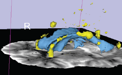
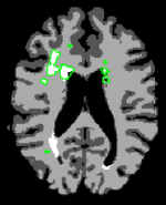
| − | | style="background:#849f66; color:#3b3f33; font-size:110%" align="Center"| <span id="1.1"></span> ''' | + | | style="background:#849f66; color:#3b3f33; font-size:110%" align="Center"| <span id="1.1"></span> '''Lupus''' |
| style="background:#CCFF99; color:black" | '''[http://www.na-mic.org/Wiki/index.php/File:Slicer3Training_WhiteMatterLesions_v2.2.1.pdf White Matter Lesions Segmentation V2.2] ''' | | style="background:#CCFF99; color:black" | '''[http://www.na-mic.org/Wiki/index.php/File:Slicer3Training_WhiteMatterLesions_v2.2.1.pdf White Matter Lesions Segmentation V2.2] ''' | ||
| style="background:#CCFF99; color:black" | [http://www.na-mic.org/Wiki/index.php/File:LesionSegmentationTutorialData.zip Lesion Segmentation Tutorial Data] | | style="background:#CCFF99; color:black" | [http://www.na-mic.org/Wiki/index.php/File:LesionSegmentationTutorialData.zip Lesion Segmentation Tutorial Data] | ||
| style="background:#CCFF99; color:black" align="Center"| [[Image:LupusTutorial2.PNG| 250px]] | | style="background:#CCFF99; color:black" align="Center"| [[Image:LupusTutorial2.PNG| 250px]] | ||
|- | |- | ||
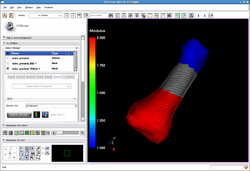
| − | | style="background:#849f66; color:#3b3f33; font-size:110%" align="Center"| <span id="1.1"></span> ''' | + | | style="background:#849f66; color:#3b3f33; font-size:110%" align="Center"| <span id="1.1"></span> '''Automated FE Mesh''' |
| style="background:#CCFF99; color:black" | '''[http://www.na-mic.org/Wiki/images/b/b8/IA-FEMesh-Tutorial-Notes.pdf IA FEMesh Tutorial] ''' | | style="background:#CCFF99; color:black" | '''[http://www.na-mic.org/Wiki/images/b/b8/IA-FEMesh-Tutorial-Notes.pdf IA FEMesh Tutorial] ''' | ||
| style="background:#CCFF99; color:black" | [http://www.na-mic.org/Wiki/index.php/File:MeshTutorialExampleData.zip Mesh Tutorial Example Data] | | style="background:#CCFF99; color:black" | [http://www.na-mic.org/Wiki/index.php/File:MeshTutorialExampleData.zip Mesh Tutorial Example Data] | ||
| style="background:#CCFF99; color:black" align="Center"| [[Image:Femesh-in-trunk-120808.png |250px]] | | style="background:#CCFF99; color:black" align="Center"| [[Image:Femesh-in-trunk-120808.png |250px]] | ||
|- | |- | ||
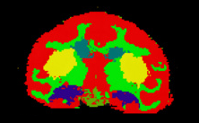
| − | | style="background:#849f66; color:#3b3f33; font-size:110%" align="Center"| <span id="1.1"></span> ''' | + | | style="background:#849f66; color:#3b3f33; font-size:110%" align="Center"| <span id="1.1"></span> '''Alcohol and Stress Interaction''' |
| style="background:#CCFF99; color:black" | '''[http://www.na-mic.org/Wiki/images/8/83/EMSegment_TrainingTutorial.pdf Non-Human Primate Segmentation Tutorial] ''' | | style="background:#CCFF99; color:black" | '''[http://www.na-mic.org/Wiki/images/8/83/EMSegment_TrainingTutorial.pdf Non-Human Primate Segmentation Tutorial] ''' | ||
| style="background:#CCFF99; color:black" | [http://www.bsl.ece.vt.edu/data/vervet_atlas/vervet.php Non-Human Primate Segmentation Data] | | style="background:#CCFF99; color:black" | [http://www.bsl.ece.vt.edu/data/vervet_atlas/vervet.php Non-Human Primate Segmentation Data] | ||
| style="background:#CCFF99; color:black" align="Center"| [[Image: Non-HumanPrimateSegmentation.png|250px]] | | style="background:#CCFF99; color:black" align="Center"| [[Image: Non-HumanPrimateSegmentation.png|250px]] | ||
|- | |- | ||
| − | | style="background:#849f66; color:#3b3f33; font-size:110%" align="Center"| <span id="1.1"></span> ''' | + | | style="background:#849f66; color:#3b3f33; font-size:110%" align="Center"| <span id="1.1"></span> '''Prostate''' |
| style="background:#CCFF99; color:black" | '''[http://wiki.na-mic.org/Wiki/index.php/IGT:ToolKit/Prostate-Planning MR Guided Prostate Interventions ]''' | | style="background:#CCFF99; color:black" | '''[http://wiki.na-mic.org/Wiki/index.php/IGT:ToolKit/Prostate-Planning MR Guided Prostate Interventions ]''' | ||
| style="background:#CCFF99; color:black" | [http://www.na-mic.org/Wiki/index.php/File:MRGuidedProstateInterventions.zip MR Guided Prostate Interventions Data] | | style="background:#CCFF99; color:black" | [http://www.na-mic.org/Wiki/index.php/File:MRGuidedProstateInterventions.zip MR Guided Prostate Interventions Data] | ||
| style="background:#CCFF99; color:black" align="Center"|[[Image:ProstatePlanningOverview.jpg | 250px]] | | style="background:#CCFF99; color:black" align="Center"|[[Image:ProstatePlanningOverview.jpg | 250px]] | ||
|- | |- | ||
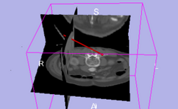
| − | | style="background:#849f66; color:#3b3f33; font-size:110%" align="Center"| <span id="1.1"></span> ''' | + | | style="background:#849f66; color:#3b3f33; font-size:110%" align="Center"| <span id="1.1"></span> '''IGT''' |
| style="background:#CCFF99; color:black" | '''[http://www.na-mic.org/Wiki/index.php/IGT:ToolKit/Navigation-tutorial Basic Navigation Tutorial] ''' | | style="background:#CCFF99; color:black" | '''[http://www.na-mic.org/Wiki/index.php/IGT:ToolKit/Navigation-tutorial Basic Navigation Tutorial] ''' | ||
| style="background:#CCFF99; color:black" | [http://www.na-mic.org/publications/item/view/1265 Basic Navigation Data] | | style="background:#CCFF99; color:black" | [http://www.na-mic.org/publications/item/view/1265 Basic Navigation Data] | ||
| style="background:#CCFF99; color:black" align="Center"| [[Image:300px-IGTBasicNavigationSummary.png| 250px]] | | style="background:#CCFF99; color:black" align="Center"| [[Image:300px-IGTBasicNavigationSummary.png| 250px]] | ||
|- | |- | ||
| − | | style="background:#849f66; color:#3b3f33; font-size:110%" align="Center"| <span id="1.1"></span> ''' | + | | style="background:#849f66; color:#3b3f33; font-size:110%" align="Center"| <span id="1.1"></span> '''Schizophrenia''' |
| style="background:#CCFF99; color:black" | '''[http://www.na-mic.org/Wiki/index.php/Python_Stochastic_Tractography_Tutorial Python Stochastic Tractography Tutorial V1.0] ''' | | style="background:#CCFF99; color:black" | '''[http://www.na-mic.org/Wiki/index.php/Python_Stochastic_Tractography_Tutorial Python Stochastic Tractography Tutorial V1.0] ''' | ||
| style="background:#CCFF99; color:black" | [http://www.na-mic.org/Wiki/index.php/File:IJdata.tar.gz Stochastic Tractography Data] | | style="background:#CCFF99; color:black" | [http://www.na-mic.org/Wiki/index.php/File:IJdata.tar.gz Stochastic Tractography Data] | ||
| style="background:#CCFF99; color:black" align="Center"| [[Image:StochasticTractographyTutorial2.png| 250px]] | | style="background:#CCFF99; color:black" align="Center"| [[Image:StochasticTractographyTutorial2.png| 250px]] | ||
|- | |- | ||
| − | | style="background:#849f66; color:#3b3f33; font-size:110%" align="Center"| <span id="1.1"></span> ''' | + | | style="background:#849f66; color:#3b3f33; font-size:110%" align="Center"| <span id="1.1"></span> '''Autism''' |
| style="background:#CCFF99; color:black" | '''[http://www.na-mic.org/Wiki/images/3/33/ARCTIC-Slicer3-Tutorial.pdf ARCTIC: Automatic Regional Cortical ThICkness V1.0]''' | | style="background:#CCFF99; color:black" | '''[http://www.na-mic.org/Wiki/images/3/33/ARCTIC-Slicer3-Tutorial.pdf ARCTIC: Automatic Regional Cortical ThICkness V1.0]''' | ||
| style="background:#CCFF99; color:black" | [http://www.nitrc.org/projects/arctic/ ARCTIC Data] | | style="background:#CCFF99; color:black" | [http://www.nitrc.org/projects/arctic/ ARCTIC Data] | ||
| style="background:#CCFF99; color:black" align="Center"| [[Image:ArcticLogo.png|250px]] | | style="background:#CCFF99; color:black" align="Center"| [[Image:ArcticLogo.png|250px]] | ||
|- | |- | ||
| − | | style="background:#849f66; color:#3b3f33; font-size:110%" align="Center"| <span id="1.1"></span> ''' | + | | style="background:#849f66; color:#3b3f33; font-size:110%" align="Center"| <span id="1.1"></span> '''Lupus''' |
| style="background:#CCFF99; color:black" | '''[http://www.na-mic.org/Wiki/index.php/File:Slicer3Training_WhiteMatterLesions_v2.0.ppt.pdf White Matter Lesions Segmentation V2.0] ''' | | style="background:#CCFF99; color:black" | '''[http://www.na-mic.org/Wiki/index.php/File:Slicer3Training_WhiteMatterLesions_v2.0.ppt.pdf White Matter Lesions Segmentation V2.0] ''' | ||
| style="background:#CCFF99; color:black" | [http://www.na-mic.org/Wiki/index.php/File:LesionSegmentationTutorialData.zip Lesion Segmentation Tutorial Data] | | style="background:#CCFF99; color:black" | [http://www.na-mic.org/Wiki/index.php/File:LesionSegmentationTutorialData.zip Lesion Segmentation Tutorial Data] | ||
| style="background:#CCFF99; color:black" align="Center"| [[Image:Lupus3.png| 150px]] | | style="background:#CCFF99; color:black" align="Center"| [[Image:Lupus3.png| 150px]] | ||
|- | |- | ||
| − | | style="background:#849f66; color:#3b3f33; font-size:110%" align="Center"| <span id="1.1"></span> ''' | + | | style="background:#849f66; color:#3b3f33; font-size:110%" align="Center"| <span id="1.1"></span> '''Orbit Segmentation''' |
| style="background:#CCFF99; color:black" | '''[[Media:OrbitSegmentationTutorial.pdf| Segmentation of the Orbit ]]''' | | style="background:#CCFF99; color:black" | '''[[Media:OrbitSegmentationTutorial.pdf| Segmentation of the Orbit ]]''' | ||
| style="background:#CCFF99; color:black" | [[Media:OrbiteSegmentationData.zip | Orbit Segmentation Data ]] | | style="background:#CCFF99; color:black" | [[Media:OrbiteSegmentationData.zip | Orbit Segmentation Data ]] | ||
| style="background:#CCFF99; color:black" align="Center"| [[Image:OrbitSegmentationt.png |250px]] | | style="background:#CCFF99; color:black" align="Center"| [[Image:OrbitSegmentationt.png |250px]] | ||
|- | |- | ||
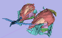
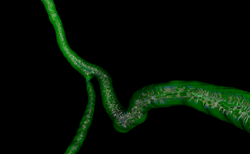
| − | | style="background:#849f66; color:#3b3f33; font-size:110%" align="Center"| <span id="1.1"></span> ''' | + | | style="background:#849f66; color:#3b3f33; font-size:110%" align="Center"| <span id="1.1"></span> '''VMTK''' |
| style="background:#CCFF99; color:black" | '''[http://www.na-mic.org/Wiki/images/4/40/TutorialVMTKCoronariesCenterlinesMRI_Winter2010AHM.pdf VMTK Coronary Arteries Centrelines Extraction]''' | | style="background:#CCFF99; color:black" | '''[http://www.na-mic.org/Wiki/images/4/40/TutorialVMTKCoronariesCenterlinesMRI_Winter2010AHM.pdf VMTK Coronary Arteries Centrelines Extraction]''' | ||
| style="background:#CCFF99; color:black" | [http://www.na-mic.org/Wiki/images/a/aa/TutorialVMTKCoronariesCenterlinesMRI_Data_Winter2010AHM.zip VMTK Centrelines Data] | | style="background:#CCFF99; color:black" | [http://www.na-mic.org/Wiki/images/a/aa/TutorialVMTKCoronariesCenterlinesMRI_Data_Winter2010AHM.zip VMTK Centrelines Data] | ||
| style="background:#CCFF99; color:black" align="Center"| [[Image:Vmtkcloseupvoronoicenterlinewithreference.png|250px]] | | style="background:#CCFF99; color:black" align="Center"| [[Image:Vmtkcloseupvoronoicenterlinewithreference.png|250px]] | ||
|- | |- | ||
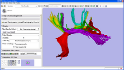
| − | | style="background:#849f66; color:#3b3f33; font-size:110%" align="Center"| <span id="1.1"></span> ''' | + | | style="background:#849f66; color:#3b3f33; font-size:110%" align="Center"| <span id="1.1"></span> '''Fiber Clustering''' |
| style="background:#CCFF99; color:black" | '''[http://www.na-mic.org/Wiki/index.php/File:FiberClusteringTrainingTutorial_Winter2010AHM.pdf| EM Fiber Clustering ]''' | | style="background:#CCFF99; color:black" | '''[http://www.na-mic.org/Wiki/index.php/File:FiberClusteringTrainingTutorial_Winter2010AHM.pdf| EM Fiber Clustering ]''' | ||
| style="background:#CCFF99; color:black" | [http://www.nitrc.org/projects/quantitativedti/ EM Fiber Clustering Data ] | | style="background:#CCFF99; color:black" | [http://www.nitrc.org/projects/quantitativedti/ EM Fiber Clustering Data ] | ||
Revision as of 14:39, 3 May 2010
Home < Slicer3.4:TrainingContents
Slicer 3.4 Tutorials
- The following table contains "How to" tutorials with matched sample data sets. They demonstrate how to use the 3D Slicer environment (version 3.4) to accomplish certain tasks.
- For questions related to the Slicer3.4 Compendium, please send an e-mail to Sonia Pujol, Ph.D. (spujol at bwh.harvard.edu).
| Category | Tutorial | Sample Data | Image |
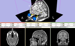
| Basic | Slicer3Minute Tutorial The Slicer3Minute tutorial is an introduction to the advanced 3D visualization capabilities of Slicer3.4. Audience: First time users. |
Slicer3Minute dataset The Slicer3Minute dataset contains an MR scan of the brain and 3D reconstructions of the anatomy |
|
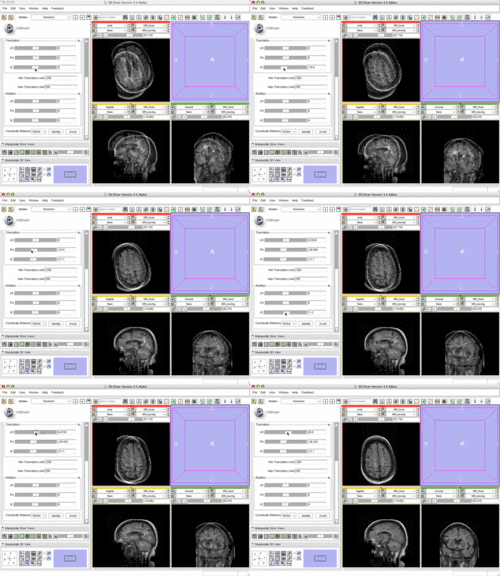
| Basic | Slicer3 Manual Registration Tutorial shows how to manually/interactively align two images in Slicer3.4 or 3.5 Audience: First time & early users. |
Manual Registration example dataset This dataset contains two brain MRI of a single subject, obtained in different orientations. |
|
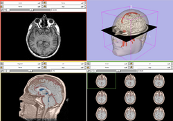
| Core | Slicer3Visualization Tutorial The Slicer3Visualization tutorial guides through 3D data loading and visualization in Slicer3.4. Audience: All beginning users including clinicians, scientists, engineers and programmers. |
Slicer3Visualization dataset The Slicer3VisualizationDataset contains two MR scans of the brain, a pre-computed labelmap and 3D reconstructions of the anatomy. |
|
| Core | Programming in Slicer3 Tutorial The Programming in Slicer3 tutorial is an introduction to the the integration of stand-alone programs outside of the Slicer3 source tree. Audience: Programmers and Engineers. |
HelloWorld Plugin The HelloWorld tutorial dataset contains an MR scan of the brain and pre-computed xml and C++ files for integrating the Hello World plug-in to Slicer3. |
|
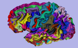
| Specialized | 3D Visualization of FreeSurfer Data The course guides through 3D visualization of FreeSurfer brain segmentations, surface reconstruction and parcellation results in Slicer3.4. Audience: All users. |
FreeSurfer Tutorial dataset The FreeSurfer dataset contains an MR scan of the brain and pre-computed FreeSurfer segmentation and cortical surface reconstructions. |
|
| Specialized | Automatic Segmentation Tutorial The course guides through the process of using the Expectation-Maximization Segmentation algorithm to automatically segment brain structures from MRI data. Audience: Programmers and Engineers. |
Automatic Segmentation dataset | 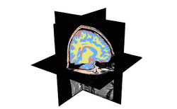
|
| Specialized | Atlas Label Merging & Surface Based Registration Tutorial This tutorial guides through the creation and co-registration of surface models of atlas structures (thalamus & thalamic nuclei) and subsequent merging of two labelmaps. |
Atlas Merging tutorial dataset (contains 2 full brain atlases + intermediate result files, 72MB) | 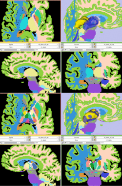
|
| Specialized | Neurosurgical Planning Tutorial This tutorial takes the trainee through a complete workup for neurosurgical patient-specific mapping. Also see this tutorial for information on how to use Slicer's affine registration, simple region growing, model maker and tractography modules. Audience: All users interested in image-guided therapy. |
Neurosurgical Planning dataset | 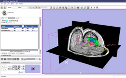
|
| Specialized | Diffusion MRI Tutorial This tutorial guides you through the process of loading diffusion MR data, estimating diffusion tensors, and performing tractography of white matter bundles. Audience: All users and developers. |
Diffusion dataset | 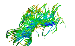
|
| Specialized | ChangeTracker Tutorial This tutorial describes the use of ChangeTracker module to detect changes in tumor volume from two MRI scans. Audience: All users interested in longitudinal analysis of pathology. |
Training data download is integrated with the ChangeTracker module (see Tutorial) | |
| Specialized | LiverSegmentation Tutorial This tutorial introduces translational clinical scientists to the capabilities of the 3D Slicer software through the segmentation of the liver. Audience: All users and developers. |
Liver Segmentation dataset | 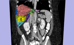
|
Slicer Tutorial Contest (under construction)
The following tutorials were part of the Summer 2009 Slicer tutorial contest and Winter 2009 Slicer tutorial contest.
Slicer 3.5 Tutorials
- The following table contains "How to" tutorials with matched sample data sets. They demonstrate how to use the 3D Slicer environment (version 3.5) to accomplish certain tasks.
| Contest | Tutorial | Sample Data | Image |
| Specialized | Robust Statistics Segmentation Tutorial | Robust Statistics Segmentation dataset | 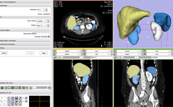
|
Software Installation
- The Slicer download page contains information on how to obtain a compiled version of Slicer for a variety of platforms and where to find the source code for Slicer 3.
Software Documentation
- For the Slicer 3.4 manual pages please click here. These pages are the reference manual for Slicer 3.4 and briefly explain the functionality found in panels and modules.
Older Tutorials
- Visit the Slicer 3.2 training page.
- Visit the Slicer 2 training page.

