File:Screen-shot of the GeodesicSlicer program. .png
From Slicer Wiki
Size of this preview: 624 × 599 pixels. Other resolutions: 250 × 240 pixels | 785 × 754 pixels.
Original file (785 × 754 pixels, file size: 199 KB, MIME type: image/png)
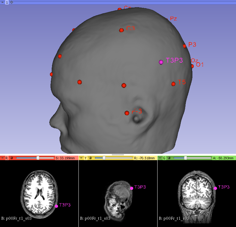
The users enters 1) the T1-weighted whole-brain anatomical image 2) Place four points: the nasion, the inion, the left tragus and the right tragus. The program make a 3D mesh morphed to the structural MRI data of a participant and calculates the 10-20 system EEG with T3P3, and outputs the distance between the anatomical target and the T3 electrode.
File history
Click on a date/time to view the file as it appeared at that time.
| Date/Time | Thumbnail | Dimensions | User | Comment | |
|---|---|---|---|---|---|
| current | 13:50, 9 March 2018 | | 785 × 754 (199 KB) | Frederic (talk | contribs) |
- You cannot overwrite this file.