Documentation/4.4/Extensions/PercutaneousApproachAnalysis
|
For the latest Slicer documentation, visit the read-the-docs. |
Introduction and Acknowledgements
|
This work is supported by Shiga University of Medical Science (SUMS) in Japan, NA-MIC, NCIGT, and Slicer Community.
This work is also supported in part by the National Institute of Health (R01CA111288, P01CA067165, P41RR019703, P41EB015898, R01CA124377, R01CA138586, R42CA137886), and was presented in CARS 2014 meetingMedia:CARS2014 Murakami.pdf. | |||||||||
|
Module Description

|
|
|
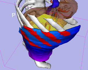
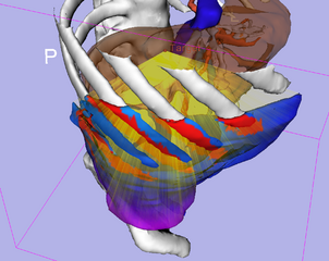
The PercutaneousApproachAnalysis module is a Slicer4 module designed to visualize bundles of needle path candidates and color map on a region in skin to avoid blood vessels, bones and specific anatomies and specify skin entry points on the path candidates. The bundles of path candidates and the skin entry point on each path in the bundles are calculated based on a target point, a skin model and an obstacle model. This bundle expression might help you grasp safety paths easily. It is also possible to evaluate the each path on the bundles based on the length and the trajectory comparing with the longest and the shortest paths. The location of the skin entry point on each path could be modified by extending and shrinking the path. The skin entry point on each path could be output as a MarkupFiducial List. After getting the outcomes, you can clean up the models and transform matrixes generated from this module. For quantitative analysis, the software calculates the ratio of the total accessible area to the total area of the Body Surface model as the accessibility score (AS) for the specified fiducial point in the liver. Additionally, the software calculates and visualizes the distance between the target tumor and each polygon of the accessible area on the Body Surface model.
Use cases
The PercutaneousPathDesigner module is a useful tool for planning needle and probe paths. Percutaneous thermal ablations including radio frequency, microwave and cryotherapy require such probe trajectories to avoid blood vessels, bones and specific anatomies to reach the probes to target tumors. Needle biopsy also requires safety paths to gather multiple tissue. For these cases, this module can help to obtain the path candidates based on the target point, a segmented obstacle model to avoid and another segmented model which represents skin entry surface.
Parameters and Outcomes
Parameters
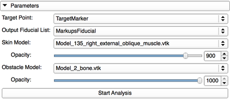
- Target Point: A fiducial point as a target of a needle insertion
- Obstacle Model: A model which you think as an obstacle of the needle insertion
- Skin Model: A model which represents a region for the candidate of the skin entry points
Outcomes
- Accessibility Score: The number calculated by accessibility area and total area on the selected Skin Model
- Color Mapped Skin: The visualization data represents the accessibility score
- All Path: The visualization data represents the candidate of the straight paths
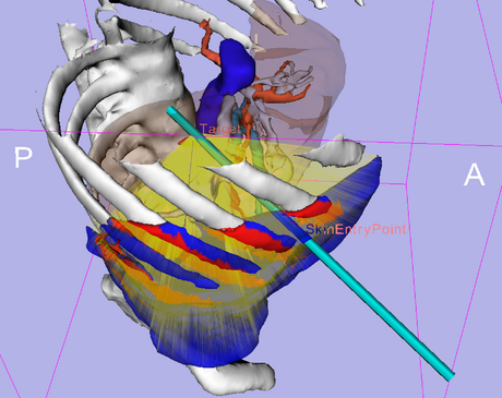
- The Longest Path: The maximum distance between the Target Point and the surface of Skin Model
- The Shortest Path: The minimum distance between the Target Point and the surface of Skin Model
- Path Candidate: All path candidate
- Skin Entry Point: Skin entry point on the selected path
Tutorials
|
- This PercutaneousApproach Analysis module could be used for the percutaneous path planning. After the analysis on this module, you can select the candidate paths from the skin to the target to avoid obstacles such as blood vessels, bones and specific anatomies and obtain the skin entry point. By using the obtained target and the skin entry points, the needle model could be placed on the module outcome by using PortPlacement module.
Tutorial steps
1. Download Tutorial Data Set: Media:PAAModuleTutorialData.zip.
2. Open "WorkBasedOnAbdominal_Atlas_2008.mrml" scene file.
3. Select "PercutaneousApproachAnalysis" module.
4. Set "F" as Target Point.
5. Set "Create new MarkupsFiducial" as Output Fiducial List.
6. Set "Model_135_right_external_oblique_muscle.vtk" as Skin Model.
7. Set "Model_2_bone.vtk" as Obstacle Model.
8. Click "Start Analysis" button.
9. Click "The shortest Path (Blue)" check box.
10. Click "Path Candidate (Red)" check box.
11. Move "Path Candidate (No.)"slider bar and make sure you can obtain each path candidate visually.
12. Set "22318" to "Path Candidate (No.)" number box manually. The number represents The shortest Path number.
13. Move "Point Candidate on the Path" slider to 600 and make sure the crossing point between the path and skin model was very close to the red color map.
14. Increment "Path Candidate (No.)" from 22318 to 22328 manually and make sure the path was going away from the red color map.
15. Change "Color Mapped Skin Opacity" to 100.
16. Move "Point Candidate on the Path" slider to 133 and make sure the crossing point between the path and skin model became a skin entry point.
17. Click "Create Point on the Path" button. You can obtain the skin entry point.
15. Select "PortPlacement" module.
16. Set "MarkupsFiducilal" as Markups node of surgical ports and click "Set Port Markup Node" button.
17. Select "Visible" column.
18. Set "F" as Surgical Target Fiducial and Click "Aim Tools at Target".
19. Set "Tool radius" to 1.50.
20. You can see the probe and the probe was far from the red color map on the skin.
Panels and their use
Parameters panel
|
- Parameters panel
- Target Point requires a MarkupsFiducial List created in Markups Module which has one target markup.
- Output Fiducial List requires a MarkupsFiducial List created in Markups Module which has no markups.
- Skin Model requires a model which represents a skin, that is a surface consists of the candidates of the needle entry points.
- Opacity Slider can change the opacity parameter for the Skin Model selected.
- Obstacle Model requires a model which represents the obstacle needle paths should avoid.
- Opacity Slider can change the opacity parameter for the Obstacle Model selected.
- Start Analysis button starts calculating path candidates from the skin model to the target point to avoid the obstacle model.
Outcomes panel
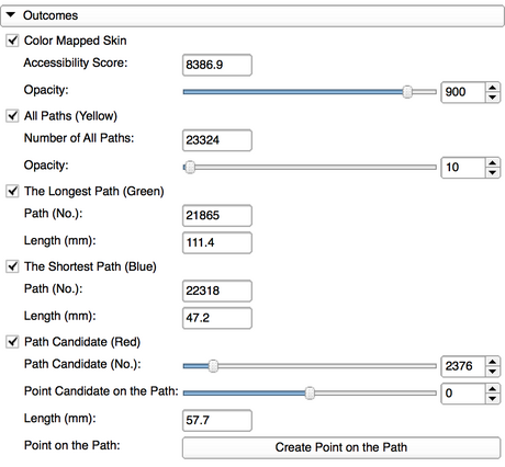
|
- Outcomes panel
- Color Mapped Skin check button visualizes a color map showing approachable and inapproachable areas on the skin model.
- Accessibility score is displayed in the result field.
- All Paths (Yellow) check button visualizes all path candidates shown as the yellow bundles of lines.
- Opacity Slider can change the opacity parameter for the yellow bundles.
- The Longest Path (Green) check button displays the longest path as the green line.
- The Shortest Path (Blue) check button displays the shortest path as the blue line.
- The Path Candidate (Red) check button displays the one of the yellow bundles as the red line.
- Path Candidate (No.) Slider can select a red line from the yellow bundles.
- Point Candidate on the Path Slider can extend and shrink the selected red line from the skin entry point.
- Create Point on the Path button adds the entry position to the Output Fiducial List you set in the Parameters panel.
Cleaning Outcomes panel

|
- Cleaning Outcomes panel
- Delete the Created Paths button erases the all models created by this module.
- Delete the Created Transforms button erases the transforms created by this module except the marker transforms.
Similar Modules
N/A
References
[1] Rossi S, Di Stasi M, Buscarini E, Cavanna L, Quaretti P, Squassante E, Garbagnati F, Buscarini L. (1995) Percutaneous radiofrequency interstitial thermal ablation in the treatment of small hepatocellular carcinoma. Cancer J Sci Am 1(1): 73-81
[2] Silverman SG, Tuncali K, Adams DF, van Sonnenberg E, Zou KH, Kacher DF, Morrison PR, Jolesz FA. (2000) MR Imaging-guided Percutaneous Cryotherapy of Liver Tumors: Initial Experience. Radiology 217: 657-64
[3] Ido K, Isoda N, Sugano K. (2001) Microwave coagulation therapy for liver cancer: laparoscopic microwave coagulation. J Gastroenterol 36(3): 145-52
[4] Khlebnikov R, Kainz B, Muehl J, Schmalstieg D. (2011) Crepscular Rays for Tumor Accessibility Planning. IEEE Trans Vis Comput Graph 17(12): 2163-72
[5] PortPlacement module (used in tutorial)
[6] SPL Abdominal Atlas (used in examples)
[7] Github source code repository (https://github.com/SNRLab/PercutaneousApproachAnalysis)
[8] Percutaneous Approach Analysis project in 2014 Winter Project Week
Logo
|
Information for Developers
| Section under construction. |

