Difference between revisions of "EMSegmenter-Tasks"
Belhachemi (talk | contribs) (Undo revision 19814 by Belhachemi (Talk)) |
Belhachemi (talk | contribs) |
||
| Line 58: | Line 58: | ||
</gallery> | </gallery> | ||
| − | ==MS Study== | + | ==Pediatric MS Study== |
| − | * Collaborator: Alexander Zaitsev, | + | * Collaborator: Alexander Zaitsev, MGH, BWH MS Center |
| − | * Short description: | + | * Short description: Evaluate brain atrophy for pediatric MS Patients |
| − | * Image specification: | + | * Image specification: 3 Tesla Scanner: SIGNA HDx / GE MEDICAL SYSTEMS, 3D MPRAGE, Sagital Scans, TR/TI/TE=24/0/7 ms, pixel_xsize = 0.976600, pixel_ysize = 0.976600, fov = 250.009598, aspect = 1.535941, thick = 1.500000, space = 0.000000 |
* Used Task: [[EMSegmenter-Tasks:MRI-Human-Brain|'''MRI Human Brain''']] | * Used Task: [[EMSegmenter-Tasks:MRI-Human-Brain|'''MRI Human Brain''']] | ||
<gallery perrow=1: widths=630px : heights=210px> | <gallery perrow=1: widths=630px : heights=210px> | ||
Revision as of 17:15, 29 April 2011
Home < EMSegmenter-TasksReturn to EMSegmenter Overview Page
Contents
What is a task?
Most clinicians are using the EMSegmenter to segment specific parts of the human body. Those segmentations depend heavily on
the used input data. The signal level in the input data varies with the used image modalities (e.g. MRI, CT, ...) and with each anatomical structure.
The EMSegmenter can take advantage of some knowledge about the signal level of those anatomical structures in the different image modalities.
Technically, each tasks consist of a .mrml file and a .tcl file.
The .mrml file stores the anatomical properties (mean values, covariance values) in a user defined tree structure.
The .tcl script is used to perform some pre-processing on the input data.
Visit the following web page to create your own task.
Segmentation Task Library
Existing Tasks in Slicer 3.6.3
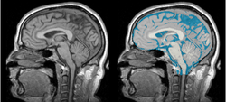 Task 01: MRI Human Brain for non-skull stripped T1 scans
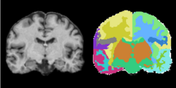 Task 02: MRI Human Brain Parcellation for skull stripped T1 scans
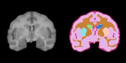 Task 03: Non-Human Primate for skull stripped T1 scans


Tasks in development for Slicer 3.6.4
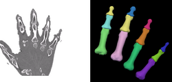 Task 04: CT Hand Bone
 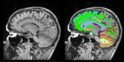 Task 05: MRI Human Brain Full Parcellation for non-skull stripped T1 scans
 Task 06: MRI Human Brain with high in-plane resolution for non-skull stripped T1 scans
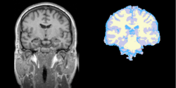 Task 07: MRI Human Brain Experimental with skull stripping
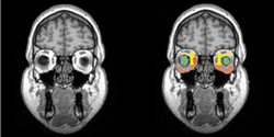 Task 08: Human Eye


EMSegmenter use cases
Quantitative assessment using MPRAGE and Flair images
- Collaborator: Tammie Benzinger , Washington University School of Medicine
- Short description: Quantitative assessment using MPRAGE and Flair images
- Image specification: Dimension: 256x256x160 , Spacing 1x1x1, T2 MPRAGE
- Used Task: MRI Human Brain Experimental (with skull stripping)
Aging Study
- Collaborator: Alexander Zaitsev, Brigham and Women's Hospital
- Short description:
- Image specification:
- Used Task: MRI Human Brain


HIV Study
- Collaborator: Alexander Zaitsev, Brigham and Women's Hospital
- Short description:
- Image specification:
- Used Task: MRI Human Brain


Pediatric MS Study
- Collaborator: Alexander Zaitsev, MGH, BWH MS Center
- Short description: Evaluate brain atrophy for pediatric MS Patients
- Image specification: 3 Tesla Scanner: SIGNA HDx / GE MEDICAL SYSTEMS, 3D MPRAGE, Sagital Scans, TR/TI/TE=24/0/7 ms, pixel_xsize = 0.976600, pixel_ysize = 0.976600, fov = 250.009598, aspect = 1.535941, thick = 1.500000, space = 0.000000
- Used Task: MRI Human Brain

Social Cognition Study
- Collaborator: Alexander Zaitsev, Brigham and Women's Hospital, Pecs University
- Short description: Evaluate brain atrophy for elderly MS patients.
- Image specification: 3 Tesla Scanner: Magnetom TIM Trio, Siemens, 3D MPRAGE, Slice thickness: 1.2mm, Spacing 1.2 1 1 (mm)
- Used Task: MRI Human Brain
