Documentation/Nightly/Extensions/BrainVolumeRefinement
|
For the latest Slicer documentation, visit the read-the-docs. |
Introduction and Acknowledgements
|
This work was partially funded by CAPES and CNPq, Brazilian Agencies. Information on CAPES can be obtained on the CAPES website and CNPq website. | |||||||||
|
Extension Description
The Brain Volume Refinement (BVeR) extension is designed to assist biomedical studies that uses MRI structural images of the healthy brain. The method is ... [1].
The BVeR algorithm is suitable for a broad use of healthy brain structural MRI images, e.g. T1w and T2w, offering broad application in many large data analyses. The main contribution of the proposed method is related to the reduction of manual interference in the brain volume refinement after an automatic skull stripping procedure been performed, helping to reduce human errors and processing time. Even though the BVeR method does not provide a fully brain extraction algorithm, it can be helpful as a ad hoc image processing step in which increase the quality of well-known brain extraction algorithm in the literature. Although not all the spectrum of skull stripping algorithm were not coverage in this study, we believe that a similar outcome can be generalized for many other brain extraction frameworks such as BEasT, 3DSkullStrip, ROBEX, OptiBET and many others. Finally, a diverse image post-processing analysis that are sensible to the brain volume estimate could also be improved by better tissue segmentation, e.g. cortical thickness and brain atrophy, which provide a valuable incentive to many biomedical studies.
Modules
- Structural T1w and T2w brain volume correction: BVeR
Use Cases
Most frequently used for these scenarios:
- Use Case 1: Noise reduction as a pre-processing step for tissue segmentation
- When dealing with single voxel classification schemes, a noise reduction pre-processing step is usually helpful to reduce data fluctuation due to acquisition artifacts (e.g. reducing the number of misclassified voxels).
- Use Case 2: Volume rendering
- Noise reduction will result in nicer looking volume renderings
- Use Case 3: Noise reduction as part of image processing pipeline
- Could offer a better segmentation and classification on specific brain image analysis such as in Multiple Sclerosis lesion segmentation

Raw T1 weighted MRI Image
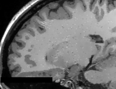
T1 weighted MRI Image with AAD filter (q=1.2)
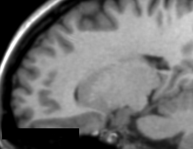
T1 weighted MRI Image with IAD filter (q=1.2)
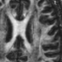
DTI-FA map without image filtering process

DTI-FA map with AAD image filtering (q=0.4)


Similar Extensions
References
- da S Senra Filho, A.C., Garrido Salmon, C.E. & Murta Junior, L.O., 2015. Anomalous diffusion process applied to magnetic resonance image enhancement. Physics in Medicine and Biology, 60(6), pp.2355–2373. DOI: 10.1088/0031-9155/60/6/2355
- Filho, A.C. da S.S. et al., 2014. Anisotropic Anomalous Diffusion Filtering Applied to Relaxation Time Estimation in Magnetic Resonance Imaging. In Annual International Conference of the IEEE Engineering in Medicine and Biology Society. IEEE, pp. 3893–3896.
- Filho, A.C. da S.S., Barizon, G.C. & Junior, L.O.M., 2014. Myocardium Segmentation Improvement with Anisotropic Anomalous Diffusion Filter Applied to Cardiac Magnetic Resonance Imaging. In Annual Meeting of Computing in Cardiology.
- Filho, A.C. da S.S. et al., 2014. Brain Activation Inhomogeneity Highlighted by the Isotropic Anomalous Diffusion Filter. In Annual International Conference of the IEEE Engineering in Medicine and Biology Society. Chicago: IEEE, pp. 3313–3316.
- Senra Filho, A.C. da S., Duque, J.J. & Murta, L.O., 2013. Isotropic anomalous filtering in Diffusion-Weighted Magnetic Resonance Imaging. I. E. in M. and B. Society, ed. Conference proceedings : ... Annual International Conference of the IEEE Engineering in Medicine and Biology Society. IEEE Engineering in Medicine and Biology Society. Conference, 2013, pp.4022–5.
Information for Developers
| Section under construction. |
Repositories:
- Source code: GitHub repository
- Issue tracker: open issues and enhancement requests
- ↑ Tsallis, C. (2009). Introduction to Nonextensive Statistical Mechanics: Approaching a Complex World. Springer.