Difference between revisions of "Documentation/Nightly/Announcements"
| Line 63: | Line 63: | ||
<gallery caption="New and Improved Extensions" widths="250px" heights="150px" perrow="4"> | <gallery caption="New and Improved Extensions" widths="250px" heights="150px" perrow="4"> | ||
| + | Image:AnglePlanes.png|[[Documentation/{{documentation/version}}/Extensions/AnglePlanesExtension|AnglePlanesExtension]] This Module is used to calculate the angle between two planes by using the normals. The user gets the choice to use two planes which are already implemented on Slicer or they can define a plane by using landmarks (at least 3 landmarks). Plane can also be saved to be reused for other models. {{updated}} | ||
| − | Image: | + | Image:CMRTK-logo.png|[[Documentation/{{documentation/version}}/Extensions/Cardiac_MRI_Toolkit|Cardiac_MRI_Toolkit]] The Cardiac MRI Toolkit includes modules for the Utah Afib DBP {{updated}} |
| + | Image:ChangeTracker_logo.png|[[Documentation/{{documentation/version}}/Extensions/ChangeTracker|ChangeTracker]] ChangeTracker is a software tool for quantification of the subtle changes in pathology. The module provides a workflow pipeline that combines user input with the medical data. As a result we provide quantitative volumetric measurements of growth/shrinkage together with the volume rendering of the tumor and color-coded visualization of the tumor growth/shrinkage. {{updated}} | ||
| + | |||
| + | Image:SlicerCIPLogo.png|[[Documentation/{{documentation/version}}/Extensions/Chest_Imaging_Platform|Chest_Imaging_Platform]] Chest Imaging Platform is an extension for quantitative CT imaging biomarkers for lung diseases. This work is funded by the National Heart, Lung, And Blood Institute of the National Institutes of Health under Award Number R01HL116931. The content is solely the responsibility of the authors and does not necessarily represent the official views of the National Institutes of Health. {{new}} | ||
| + | |||
| + | Image:CurveMakerIcon.png|[[Documentation/{{documentation/version}}/Extensions/CurveMaker|CurveMaker]] This is an module to generate a curve based on a list of fiducial points. {{updated}} | ||
| + | |||
| + | Image:DiceSimilarityCoefficient.png|[[Documentation/{{documentation/version}}/Extensions/DiceComputation|DiceComputation]] Compute Dice's Similarity Coefficient (DSC) for several registered label map images. {{updated}} | ||
| + | |||
| + | Image:screenshot.php?group_id=403&screenshot_id=606|[[Documentation/{{documentation/version}}/Extensions/DTIAtlasFiberAnalyzer|DTIAtlasFiberAnalyzer]] DTIAtlasFiberAnalyzer allows the user to study the behavior of water diffusion (using DTI data) along the length of the white matter fiber-tracts. {{updated}} | ||
| + | |||
| + | Image:index.php?title=Main_Page|[[Documentation/{{documentation/version}}/Extensions/Eigen|Eigen]] a C++ template library for linear algebra: matrices, vectors, numerical solvers, and related algorithms. {{updated}} | ||
| + | |||
| + | Image:screenshot.php?group_id=534&screenshot_id=767|[[Documentation/{{documentation/version}}/Extensions/FiberViewerLight|FiberViewerLight]] FiberViewerLight is an open-source software to visualize and edit fibers {{updated}} | ||
| + | |||
| + | Image:GelDosimetry_Logo_128x128.png|[[Documentation/{{documentation/version}}/Extensions/GelDosimetryAnalysis|GelDosimetryAnalysis]] Slicelet covering the gel dosimetry analysis workflow used in commissioning new radiation techniques and to validate the accuracy of radiation treatment by enabling visual comparison of the planned dose to the delivered dose, where correspondence between the two dose distributions is achieved using embedded landmarks. Gel dosimetry is based on imaging chemical systems spatially fixed in gelatin, which exhibit a detectable change upon irradiation. {{updated}} | ||
| + | |||
| + | Image:GraphCutSegment.png|[[Documentation/{{documentation/version}}/Extensions/GraphCutSegment|GraphCutSegment]] This is a segment extension using graph cut and star shape algorithm. This extension can be used for research purposes ONLY. It can NOT be used for commercial purposes. {{new}} | ||
| + | |||
| + | Image:IASEM.png|[[Documentation/{{documentation/version}}/Extensions/IASEM|IASEM]] {{updated}} | ||
| + | |||
| + | Image:LightWeightRobotIGT.png|[[Documentation/{{documentation/version}}/Extensions/LightWeightRobotIGT|LightWeightRobotIGT]] 3D Slicer module for communication between 3D Slicer and LightWeight robot. {{updated}} | ||
| + | |||
| + | Image:LongitudinalPETCTLogo.png|[[Documentation/{{documentation/version}}/Extensions/LongitudinalPETCT|LongitudinalPETCT]] The purpose of the Longitudinal PET/CT Analysis module is to provide a user friendly Slicer interface for quantification of DICOM PET/CT image data by computing the standardized uptake value (SUV) based on bodyweight for different regions of interest and for different timepoints. {{updated}} | ||
| + | |||
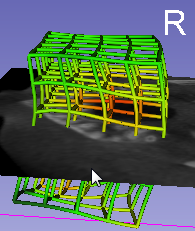
| + | Image:LumpNav.png|[[Documentation/{{documentation/version}}/Extensions/LumpNav|LumpNav]] Breast tumor resection using tracked ultrasound and cautery {{new}} | ||
| + | |||
| + | Image:MABMIS_Icon.png|[[Documentation/{{documentation/version}}/Extensions/MABMIS|MABMIS]] Multi-Atlas Based Group Segmentation {{updated}} | ||
| + | |||
| + | Image:MarginCalculator_Logo2_128.png|[[Documentation/{{documentation/version}}/Extensions/MarginCalculator|MarginCalculator]] The Matlab Bridge extension allows running Matlab scripts as command-line interface (CLI) modules directly from 3D Slicer. The only prerequisites for running Matlab scripts are having this extension and Matlab installed on the 3D Slicer computer (building of 3D Slicer, MEX files, etc. is not needed). Extension version: 0.13.0. {{updated}} | ||
| + | |||
| + | Image:MatlabBridgeLogo.png|[[Documentation/{{documentation/version}}/Extensions/MatlabBridge|MatlabBridge]] The Matlab Bridge extension allows running Matlab scripts as command-line interface (CLI) modules directly from 3D Slicer. The only prerequisites for running Matlab scripts are having this extension and Matlab installed on the 3D Slicer computer (building of 3D Slicer, MEX files, etc. is not needed). {{updated}} | ||
| + | |||
| + | Image:Slicer4ExtensionModelToModelDistance.png|[[Documentation/{{documentation/version}}/Extensions/ModelToModelDistance|ModelToModelDistance]] This extension computes the distance between two 3D models {{updated}} | ||
| + | |||
| + | Image:NeedleFinder.png|[[Documentation/{{documentation/version}}/Extensions/NeedleFinder|NeedleFinder]] NeedleFinder: fast interactive needle detection. It provides interactive tools to segment needles in MR/CT images. It has been mostly tested on MRI from gynelogical brachytherapy cases. Cf <<Validation of Catheter Segmentation for MR-Guided Gynecologic Cancer Brachytherapy.>> MICCAI 2013 {{updated}} | ||
| + | |||
| + | Image:PAAlogo-small.png|[[Documentation/{{documentation/version}}/Extensions/PercutaneousApproachAnalysis|PercutaneousApproachAnalysis]] The Percutaneous Approach Analysis is used to calculate and visualize the accessibility of liver tumor with a percutaneous approach. {{updated}} | ||
| + | |||
| + | Image:PET-IndiC.png|[[Documentation/{{documentation/version}}/Extensions/PET-IndiC|PET-IndiC]] The PET-IndiC Extension allows for fast segmentation of regions of interest and calculation of quantitative indices. {{updated}} | ||
| + | |||
| + | Image:DPetBrainQuantification.png|[[Documentation/{{documentation/version}}/Extensions/PetSpectAnalysis|PetSpectAnalysis]] First Version of the Pet Spect Analysis Extension {{updated}} | ||
| + | |||
| + | Image:PickAndPaint.png|[[Documentation/{{documentation/version}}/Extensions/PickAndPaintExtension|PickAndPaintExtension]] Pick 'n Paint tool allows users to select ROIs on a reference model and to propagate it over different time point models. {{updated}} | ||
| + | |||
| + | Image:PkModeling.png|[[Documentation/{{documentation/version}}/Extensions/PkModeling|PkModeling]] PkModeling is a Slicer4 Extension that provides pharmacokinetic modeling for dynamic contrast enhanced MRI (DCE MRI). {{updated}} | ||
| + | |||
| + | Image:Portplacement_icon.png|[[Documentation/{{documentation/version}}/Extensions/PortPlacement|PortPlacement]] Assists in the planning of surgical port placement in a laparoscopic procedure. {{updated}} | ||
| + | |||
| + | Image:ReportingLogo.png|[[Documentation/{{documentation/version}}/Extensions/Reporting|Reporting]] The purpose of the Reporting module is to provide Slicer interface for creating image annotations/markup that are stored in a structured form, and can be exported into Annotation Image Markup (AIM) XML-based format. The documentation is available at this location: http://wiki.slicer.org/slicerWiki/index.php/Documentation/4.2/Extensions/Reporting {{updated}} | ||
| + | |||
| + | Image:screenshot.php?group_id=759&screenshot_id=704|[[Documentation/{{documentation/version}}/Extensions/ShapePopulationViewer|ShapePopulationViewer]] Visualize and interact with multiple surfaces at the same time to easily compare them {{updated}} | ||
| + | |||
| + | Image:SkullStripper.png|[[Documentation/{{documentation/version}}/Extensions/SkullStripper|SkullStripper]] Skull stripping for structural MR images of the brain, tested on T1 and T2 contrast. {{updated}} | ||
| + | |||
| + | Image:AirwaySegmentation-ICON.png|[[Documentation/{{documentation/version}}/Extensions/Slicer-AirwaySegmentation|Slicer-AirwaySegmentation]] CLI module for airway segmentation of chest CT images {{updated}} | ||
| + | |||
| + | Image:SlicerHeart_Logo_128x128.png|[[Documentation/{{documentation/version}}/Extensions/SlicerHeart|SlicerHeart]] Modules for cardiac analysis and intervention planning and guidance {{new}} | ||
| + | |||
| + | Image:SlicerIGTLogo.png|[[Documentation/{{documentation/version}}/Extensions/SlicerIGT|SlicerIGT]] This extension contains modules that enable rapid prototyping of applications for image-guided interventions. Intended users should have real-time imaging and/or tracking hardware (e.g. tracked ultrasound) connected to 3D Slicer through OpenIGTLink network. Specific modules allow patient registration to the navigation coordinate system in 3D Slicer, and real-time update of tracked models and images. {{updated}} | ||
| + | |||
| + | Image:SlicerProstate_Logo_1.0_128x128.png|[[Documentation/{{documentation/version}}/Extensions/SlicerProstate|SlicerProstate]] SlicerProstate extension hosts various modules to facilitate processing and management of prostate image data, utilizing prostate images in image-guided interventions and development of the imaging biomarkers of the prostate cancer. {{new}} | ||
| + | |||
| + | Image:SlicerRT_Logo_3.0_128x128.png|[[Documentation/{{documentation/version}}/Extensions/SlicerRT|SlicerRT]] Modules for radiation therapy research. Features include DICOM-RT import/export, dose volume histogram, dose accumulation, structure comparison and morphology, isodose line/surface generation, etc. Version 0.17.1 {{updated}} | ||
| + | |||
| + | Image:Slicer-Wasp.png|[[Documentation/{{documentation/version}}/Extensions/Slicer-Wasp|Slicer-Wasp]] A module to perform a series of ITK watershed segmentation (without seeds) and then let the user create a label map out of selected components. {{new}} | ||
| + | |||
| + | Image:T1_Mapping_Logo_Resized.png|[[Documentation/{{documentation/version}}/Extensions/T1Mapping|T1Mapping]] T1 mapping estimates effective tissue parameter maps (T1) from multi-spectral FLASH MRI scans with different flip angles. {{new}} | ||
| + | |||
| + | Image:UKF_icon.png|[[Documentation/{{documentation/version}}/Extensions/UKFTractography|UKFTractography]] A framework which uses an unscented Kalman filter for performing tractography. The development of this module was supported by NIH grants R01 MH097979 (PI Rathi), R01 MH092862 (PIs Westin and Verma), U01 NS083223 (PI Westin), R01 MH074794 (PI Westin) and P41 EB015902 (PI Kikinis). {{updated}} | ||
| + | |||
| + | Image:VolumeClipLogo.png|[[Documentation/{{documentation/version}}/Extensions/VolumeClip|VolumeClip]] Clip volumes with surface models and ROI boxes {{updated}} | ||
| + | |||
| + | Image:WindowLevelEffectLogo.png|[[Documentation/{{documentation/version}}/Extensions/WindowLevelEffect|WindowLevelEffect]] Use this tool to change window/level of background/foreground volumes based on the intensity range of the selected rectangle. Normal mode: use primary mouse button to adjust window level. Depending on the selection, Foreground, Background or both layers are affected. Rectangle mode: Left Click and Drag: sweep out an outline that will draw when the button is released. The outline will define the rectangle for calculating the new window/level settings for the selected layers. {{updated}} | ||
Revision as of 08:42, 29 October 2015
Home < Documentation < Nightly < Announcements
|
For the latest Slicer documentation, visit the read-the-docs. |
 |
| Summary | What is 3D Slicer | Slicer Nightly Highlights | Slicer Training | Slicer Extensions | Other Improvements, Additions & Documentation |
Summary
The community of Slicer developers is proud to announce the release of Slicer Nightly.
- Slicer Nightly introduces
- An improved App Store, known as the Extension Manager, for adding plug-ins to Slicer. More than 80 plug-ins and packages of plug-ins are currently available.
- Close to 150 feature improvements and bug fixes have resulted in improved performance and stability.
- Improvements to many modules.
- Click here to download Slicer Nightly for different platforms and find pointers to the source code, mailing lists and the bug tracker.
- Please note that Slicer continues to be a research package and is not intended for clinical use. Testing of functionality is an ongoing activity with high priority, however, some features of Slicer are not fully tested.
- The Slicer Training page provides a series of tutorials and data sets for training in the use of Slicer.
slicer.org is the portal to the application, training materials, and the development community.
What is 3D Slicer
3D Slicer is:
- A software platform for the analysis (including registration and interactive segmentation) and visualization (including volume rendering) of medical images and for research in image guided therapy.
- A free, open source software available on multiple operating systems: Linux, MacOSX and Windows
- Extensible, with powerful plug-in capabilities for adding algorithms and applications.
Features include:
- Multi organ: from head to toe.
- Support for multi-modality imaging including, MRI, CT, US, nuclear medicine, and microscopy.
- Bidirectional interface for devices.
There is no restriction on use, but Slicer is not approved for clinical use and intended for research. Permissions and compliance with applicable rules are the responsibility of the user. For details on the license see here
Slicer Nightly Highlights
- New and Improved Modules
 Improved NameOfModule module with XXX for YYYY - click here for demo video.
- Improved ....
- Something else
- And a last one

Slicer Training
The Slicer Training page provides a series of updated tutorials and data sets for training in the use of Slicer Nightly.
- New Tutorials

This is an example of description NEW

Slicer Extensions
- New and Improved Extensions
- AnglePlanes.png
AnglePlanesExtension This Module is used to calculate the angle between two planes by using the normals. The user gets the choice to use two planes which are already implemented on Slicer or they can define a plane by using landmarks (at least 3 landmarks). Plane can also be saved to be reused for other models. UPDATED

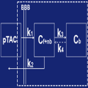
Cardiac_MRI_Toolkit The Cardiac MRI Toolkit includes modules for the Utah Afib DBP UPDATED
ChangeTracker ChangeTracker is a software tool for quantification of the subtle changes in pathology. The module provides a workflow pipeline that combines user input with the medical data. As a result we provide quantitative volumetric measurements of growth/shrinkage together with the volume rendering of the tumor and color-coded visualization of the tumor growth/shrinkage. UPDATED
- SlicerCIPLogo.png
Chest_Imaging_Platform Chest Imaging Platform is an extension for quantitative CT imaging biomarkers for lung diseases. This work is funded by the National Heart, Lung, And Blood Institute of the National Institutes of Health under Award Number R01HL116931. The content is solely the responsibility of the authors and does not necessarily represent the official views of the National Institutes of Health. NEW

CurveMaker This is an module to generate a curve based on a list of fiducial points. UPDATED

DiceComputation Compute Dice's Similarity Coefficient (DSC) for several registered label map images. UPDATED
- Screenshot.php?group id=403&screenshot id=606
DTIAtlasFiberAnalyzer DTIAtlasFiberAnalyzer allows the user to study the behavior of water diffusion (using DTI data) along the length of the white matter fiber-tracts. UPDATED
- Index.php?title=Main Page
Eigen a C++ template library for linear algebra: matrices, vectors, numerical solvers, and related algorithms. UPDATED
- Screenshot.php?group id=534&screenshot id=767
FiberViewerLight FiberViewerLight is an open-source software to visualize and edit fibers UPDATED

GelDosimetryAnalysis Slicelet covering the gel dosimetry analysis workflow used in commissioning new radiation techniques and to validate the accuracy of radiation treatment by enabling visual comparison of the planned dose to the delivered dose, where correspondence between the two dose distributions is achieved using embedded landmarks. Gel dosimetry is based on imaging chemical systems spatially fixed in gelatin, which exhibit a detectable change upon irradiation. UPDATED
GraphCutSegment This is a segment extension using graph cut and star shape algorithm. This extension can be used for research purposes ONLY. It can NOT be used for commercial purposes. NEW

IASEM UPDATED

LightWeightRobotIGT 3D Slicer module for communication between 3D Slicer and LightWeight robot. UPDATED
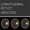
LongitudinalPETCT The purpose of the Longitudinal PET/CT Analysis module is to provide a user friendly Slicer interface for quantification of DICOM PET/CT image data by computing the standardized uptake value (SUV) based on bodyweight for different regions of interest and for different timepoints. UPDATED
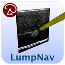
LumpNav Breast tumor resection using tracked ultrasound and cautery NEW

MABMIS Multi-Atlas Based Group Segmentation UPDATED

MarginCalculator The Matlab Bridge extension allows running Matlab scripts as command-line interface (CLI) modules directly from 3D Slicer. The only prerequisites for running Matlab scripts are having this extension and Matlab installed on the 3D Slicer computer (building of 3D Slicer, MEX files, etc. is not needed). Extension version: 0.13.0. UPDATED

MatlabBridge The Matlab Bridge extension allows running Matlab scripts as command-line interface (CLI) modules directly from 3D Slicer. The only prerequisites for running Matlab scripts are having this extension and Matlab installed on the 3D Slicer computer (building of 3D Slicer, MEX files, etc. is not needed). UPDATED
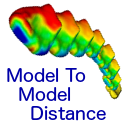
ModelToModelDistance This extension computes the distance between two 3D models UPDATED

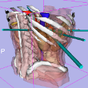
NeedleFinder NeedleFinder: fast interactive needle detection. It provides interactive tools to segment needles in MR/CT images. It has been mostly tested on MRI from gynelogical brachytherapy cases. Cf <<Validation of Catheter Segmentation for MR-Guided Gynecologic Cancer Brachytherapy.>> MICCAI 2013 UPDATED
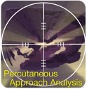
PercutaneousApproachAnalysis The Percutaneous Approach Analysis is used to calculate and visualize the accessibility of liver tumor with a percutaneous approach. UPDATED
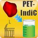
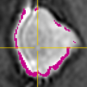
PET-IndiC The PET-IndiC Extension allows for fast segmentation of regions of interest and calculation of quantitative indices. UPDATED
PetSpectAnalysis First Version of the Pet Spect Analysis Extension UPDATED
- PickAndPaint.png
PickAndPaintExtension Pick 'n Paint tool allows users to select ROIs on a reference model and to propagate it over different time point models. UPDATED
PkModeling PkModeling is a Slicer4 Extension that provides pharmacokinetic modeling for dynamic contrast enhanced MRI (DCE MRI). UPDATED
PortPlacement Assists in the planning of surgical port placement in a laparoscopic procedure. UPDATED

Reporting The purpose of the Reporting module is to provide Slicer interface for creating image annotations/markup that are stored in a structured form, and can be exported into Annotation Image Markup (AIM) XML-based format. The documentation is available at this location: http://wiki.slicer.org/slicerWiki/index.php/Documentation/4.2/Extensions/Reporting UPDATED
- Screenshot.php?group id=759&screenshot id=704
ShapePopulationViewer Visualize and interact with multiple surfaces at the same time to easily compare them UPDATED
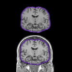
SkullStripper Skull stripping for structural MR images of the brain, tested on T1 and T2 contrast. UPDATED
Slicer-AirwaySegmentation CLI module for airway segmentation of chest CT images UPDATED

SlicerHeart Modules for cardiac analysis and intervention planning and guidance NEW
SlicerIGT This extension contains modules that enable rapid prototyping of applications for image-guided interventions. Intended users should have real-time imaging and/or tracking hardware (e.g. tracked ultrasound) connected to 3D Slicer through OpenIGTLink network. Specific modules allow patient registration to the navigation coordinate system in 3D Slicer, and real-time update of tracked models and images. UPDATED
SlicerProstate SlicerProstate extension hosts various modules to facilitate processing and management of prostate image data, utilizing prostate images in image-guided interventions and development of the imaging biomarkers of the prostate cancer. NEW

SlicerRT Modules for radiation therapy research. Features include DICOM-RT import/export, dose volume histogram, dose accumulation, structure comparison and morphology, isodose line/surface generation, etc. Version 0.17.1 UPDATED
Slicer-Wasp A module to perform a series of ITK watershed segmentation (without seeds) and then let the user create a label map out of selected components. NEW
T1Mapping T1 mapping estimates effective tissue parameter maps (T1) from multi-spectral FLASH MRI scans with different flip angles. NEW
- UKF icon.png
UKFTractography A framework which uses an unscented Kalman filter for performing tractography. The development of this module was supported by NIH grants R01 MH097979 (PI Rathi), R01 MH092862 (PIs Westin and Verma), U01 NS083223 (PI Westin), R01 MH074794 (PI Westin) and P41 EB015902 (PI Kikinis). UPDATED

VolumeClip Clip volumes with surface models and ROI boxes UPDATED
WindowLevelEffect Use this tool to change window/level of background/foreground volumes based on the intensity range of the selected rectangle. Normal mode: use primary mouse button to adjust window level. Depending on the selection, Foreground, Background or both layers are affected. Rectangle mode: Left Click and Drag: sweep out an outline that will draw when the button is released. The outline will define the rectangle for calculating the new window/level settings for the selected layers. UPDATED


Extensions removed
- NA
Extensions renamed
- PyDevRemoteDebug -> DebuggingTools
- MultidimData -> Sequences
- TrackerStabilizer -> Slicer-TrackerStabilizer
- AirwaySegmentation -> Slicer-AirwaySegmentation
Other Improvements, Additions & Documentation
To be done
For Developers
Under the hood
To be done
- Build-system
- Improved support for Visual Studio 2013
- Improved Toolkits

Moved from CTKAppLauncher v0.1.11 to v0.1.14 (43 commits)

Moved from ITK v4.4.1 to v4.6.0 (1089 commits)
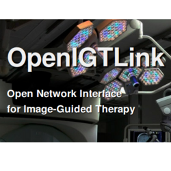
Moved from OpenIGTLink 66e272d to 849b434 (53 commits)

Moved from Qt 4.7.4 to Qt 4.8.6

Moved from VTK v5.10.1 to VTK v6.2.0 (5490 commits)
Looking at the Code Changes
From a git checkout you can easily see the all the commits since the time of the 4.4.0 release:
git log v4.3.0..HEAD
To see a summary of your own commits, you could use something like:
git log v4.3.0..HEAD --oneline --author=pieper
see the git log man page for more options.
Commit stats and full changelog