Difference between revisions of "Documentation/4.2/Training"
From Slicer Wiki
| Line 156: | Line 156: | ||
* Author: Justin Kirby | * Author: Justin Kirby | ||
*Audience: Novice 3D Slicer users interested in visualizing prostate segmentations hosted on TCIA. | *Audience: Novice 3D Slicer users interested in visualizing prostate segmentations hosted on TCIA. | ||
| − | *Summary: 3D Slicer was used to generate NRRD segmentations of the following prostate components: prostate gland boundary; internal capsule; central gland, peripheral zone; seminal vesicles; urethra; cancer – dominant nodule; neurovascular bundle; penile bulb; ejaculatory duct; veru-montanum; rectum. These segmentations are currently available for [https://wiki.cancerimagingarchive.net/display/Public/Prostate-Diagnosis 5 subjects from TCIA’s Prostate-Diagnosis image collection]. | + | *Summary: 3D Slicer was used to generate NRRD segmentations of the following prostate components: prostate gland boundary; internal capsule; central gland, peripheral zone; seminal vesicles; urethra; cancer – dominant nodule; neurovascular bundle; penile bulb; ejaculatory duct; veru-montanum; rectum. These segmentations are currently available for [https://wiki.cancerimagingarchive.net/display/Public/Prostate-Diagnosis 5 subjects from TCIA’s Prostate-Diagnosis image collection]. The [[media:File:ProstateDx-01-0006-1.zip |archive]] contains both the DICOM images and the NRRD file for one of the 5 available subjects. |
|align="right"|[[Image:NCIA-prostate.png|250px]] | |align="right"|[[Image:NCIA-prostate.png|250px]] | ||
|- | |- | ||
Revision as of 22:44, 17 December 2012
Home < Documentation < 4.2 < TrainingContents
Introduction: Slicer 4.2 Tutorials
- This page contains "How to" tutorials with matched sample data sets. They demonstrate how to use the 3D Slicer environment (version 4.2 release) to accomplish certain tasks.
- For tutorials for other versions of Slicer, please visit the Slicer training portal.
- For "reference manual" style documentation, please visit the Slicer 4.2 documentation page
- For questions related to the Slicer4 Compendium, please send an e-mail to Sonia Pujol, Ph.D
General Introduction
Slicer Welcome Tutorial
|
Slicer4Minute Tutorial
|
Slicer4 Data Loading and 3D Visualization
|
Tutorials for software developers
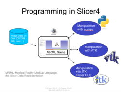
Slicer4 Programming Tutorial
|
Specific functions
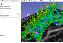
Slicer4 Diffusion Tensor Imaging Tutorial
|
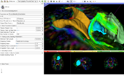
Slicer4 Neurosurgical Planning Tutorial
|
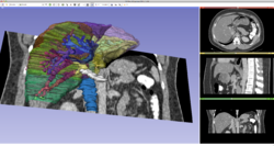
Slicer4 3D Visualization of DICOM images for Radiology Applications
|
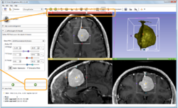
Slicer4 Quantitative Imaging tutorial
|
Summer 2012 Tutorial contest
Automatic Left Atrial Scar Segmenter
|
Qualitative and quantitative comparison of two RT dose distributions
|
Dose accumulation for adaptive radiation therapy
|
WebGL Export
|
OpenIGTLink
|
Additional resources
|
|
|
|
|
|
|
|
External Resources
Using the Editor
This set of tutorials about the use of slicer in paleontology is very well written and provides step-by-step instructions. Even though it covers slicer version 3.4, many of the concepts and techniques have applicability to the new version and to any 3D imaging field:
- Open Source Paleontologist: 3D Slicer: The Tutorial
- Open Source Paleontologist: 3D Slicer: The Tutorial Part II
- Open Source Paleontologist: 3D Slicer: The Tutorial Part III
- Open Source Paleontologist: 3D Slicer: The Tutorial Part IV
- Open Source Paleontologist: 3D Slicer: The Tutorial Part V
- Open Source Paleontologist: 3D Slicer: The Tutorial Part VI
User Contributions
See the User Contributions Page for more content.
