Documentation/Nightly/Modules/ROBEXBrainExtraction
|
For the latest Slicer documentation, visit the read-the-docs. |
Introduction and Acknowledgements
|
This work was partially funded by CAPES and CNPq (grant 201871/2015-7/SWE), a Brazilian research support agency. Information on CAPES can be obtained from the official websites, CAPES here and CAPES here. | |||||||||
|
Module Description
This module aims to extract the brain in structural MRI images, namely T1 weighted MRI images. The method is based on the non-parametric algorithm proposed by Iglesias et. al. [1]. The application with T2 and proton density MRI images were not intensively tested, but it is assumed that the algorithm is robust with those image contrasts as well. For more details about the brain extraction method, please check the original paper and also the module repository in order to see the licenses applied in this project.
In addition, for better brain volume extraction results it could be applied some preprocessing steps such as a noise attenuation and field inhomogeneity correction methods in advance. See the modules AAD Image Filter and N4 Bias Field Correction.
Use Cases
Most frequently used for these scenarios:
- Use Case 1: Initial image processing step for many neuroimage analysis, such as in DTI and cortical thickness estimation.
- It would be useful for tissue classification.
- Use Case 2: Information reduction to registration pipelines
- The non-brain tissues tend to decrease the quality of many registration algorithms.
- Use Case 3: Increase the precision of classification algorithms for neurodegenerative brain diseases diagnosis.
- Could offer a better segmentation and classification on specific brain image analysis such as in Multiple Sclerosis lesion segmentation as well as the global brain atrophy.
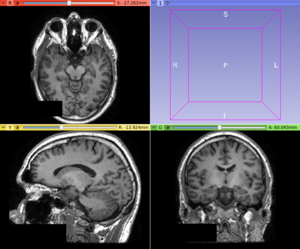
T1w MRI
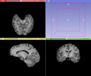
Brain extracted T1 MRI


Tutorials
N/A
Panels and their use

- Input Volume:
- Insert an input scalar volume
- Output Volume:
- Set the output volume
- Brain Mask:
- Set, if desired, the output binary mask resulted from the skull stripping process.
NOTE: The ROBEX brain extraction method is fully automatic and does not require any input parameter.
Similar Modules
References
- Iglesias JE, Liu CY, Thompson P, Tu Z: "Robust Brain Extraction Across Datasets and Comparison with Publicly Available Methods", IEEE Transactions on Medical Imaging, 30(9), 2011, 1617-1634.
Repositories:
- Official ROBEX repository: https://www.nitrc.org/projects/robex
- Source code: GitHub repository
- Issue tracker: open issues and enhancement requests
- ↑ Iglesias, J.E. et al., 2011. "Robust Brain Extraction Across Datasets and Comparison With Publicly Available Methods". IEEE Transactions on Medical Imaging, 30(9), pp.1617–1634