Uncategorized files
From Slicer Wiki
Showing below up to 500 results in range #501 to #1,000.
View (previous 500 | next 500) (20 | 50 | 100 | 250 | 500)
 AstroBeam.png 1,264 × 881; 370 KB
 AstroBlankingSegmentation1.png 1,854 × 1,010; 327 KB
 AstroBlankingSegmentation2.png 1,854 × 1,011; 287 KB
 AstroContourEffect.png 1,853 × 977; 429 KB
 AstroCroppingROI1.png 1,854 × 1,010; 333 KB
 AstroCroppingROI2.png 1,854 × 1,010; 321 KB
 AstroData.png 623 × 835; 88 KB
 AstroDataSample.png 594 × 695; 93 KB
 AstroDisplay.png 1,854 × 1,020; 312 KB
 AstroHistogram.png 1,854 × 981; 400 KB
 AstroLoading.png 1,021 × 205; 28 KB
 AstroMask2.png 1,854 × 977; 352 KB
 AstroMask3.png 1,853 × 1,020; 420 KB
 AstroMask4.png 1,854 × 1,023; 370 KB
 AstroMask6.png 1,854 × 974; 372 KB
 AstroModelingInput.png 1,856 × 1,058; 407 KB
 AstroModelingOutput.png 1,853 × 976; 447 KB
 AstroMomentMaps.png 1,854 × 1,023; 251 KB
 AstroPVDiagram.png 1,854 × 975; 1.39 MB
 AstroPVSlice.png 1,853 × 983; 375 KB
 AstroProfiles.png 1,852 × 975; 311 KB
 AstroRendering.png 1,852 × 973; 490 KB
 AstroReproject.png 1,854 × 975; 485 KB
 AstroReprojectContours.png 1,854 × 975; 752 KB
 AstroSmoothing.png 1,854 × 1,056; 496 KB
 AstroStatisticsROI.png 1,853 × 983; 293 KB
 AstroStatisticsSegmentation.png 1,854 × 1,010; 311 KB
 AstroVolumeInformation.png 1,854 × 1,022; 323 KB
 Astromed1-mj.jpg 486 × 196; 16 KB
 At sign.svg 145 × 145; 219 bytes
 Atlas-2007-05-16.png 2,469 × 1,578; 1.07 MB
 Atlas.png 599 × 600; 231 KB
 AtlasCreator-screenshot000.png 1,281 × 891; 469 KB
 AtlasCreator-screenshot001.png 1,407 × 801; 99 KB
 AtlasCreator-screenshot002.png 1,435 × 829; 118 KB
 AtlasCreatorClassDiagramm.png 1,854 × 1,326; 250 KB
 AtlasDBSyncGUI.jpg 635 × 491; 166 KB
 AtlasLabelFusion.png 451 × 279; 96 KB
 Atlas based seeding.tar.gz ; 10.6 MB
 Attention niels epting.svg 80 × 80; 4 KB
 Automated port placement surgical plan.png 830 × 674; 198 KB
- AutomaticSegmentation.zip ; 22.37 MB
 AutomaticSegmentation SoniaPujol.pdf 1,650 × 1,275, 99 pages; 12.76 MB
 Avery-Neuroimage2014-fig3.png 1,000 × 350; 433 KB
 Avf 3d centerlines.png 483 × 440; 2 KB
 Avf 3d eikonal.png 482 × 440; 68 KB
 Avf 3d evolution new.png 483 × 440; 45 KB
 Avf 3d initialization.png 483 × 440; 35 KB
 Avf 3d preparation.png 483 × 440; 39 KB
 Avf 3d voronoi big.png 815 × 504; 227 KB
 Avf fm label map.png 482 × 440; 70 KB
 Avf frangi multiscale.png 482 × 440; 23 KB
 Avf gac 20it -100 0 -100.png 482 × 440; 68 KB
 Avf gac 20it 0 0 -100.png 482 × 440; 69 KB
 Avf gac 20it 0 100 0.png 482 × 440; 70 KB
 Avf gac 20it 0 60 -100.png 482 × 440; 70 KB
 Avf orig axial.png 482 × 440; 67 KB
- Avf voismall.nrrd ; 144 KB
 Avonex.jpg 766 × 981; 631 KB
 Axer-ComputMedImagingGraph2002.pdf 1,275 × 1,754, 6 pages; 671 KB
 AxialSliceReformat.png 560 × 537; 127 KB
 Azizi-fig4.jpg 770 × 409; 199 KB
 B-spline-reg-detail.png 659 × 401; 117 KB
 B-spline-reg.png 1,356 × 1,031; 572 KB
 B.gif 600 × 459; 38.74 MB
 B2A2.png 739 × 496; 52 KB
 BC1 DCE.png 581 × 447; 150 KB
 BC1 Ktrans map ROI.png 579 × 447; 156 KB
 BCAnalysis Perfusion1.png 1,318 × 970; 296 KB
 BCCPanel.jpg 529 × 460; 29 KB
- BIRNFIPS2Slicer3-2006-10-24.ppt ; 1.4 MB
 BIS-Slicer-2009-08-13.jpg 1,216 × 980; 107 KB
 BRAINSDemonWarpForm.png 397 × 750; 24 KB
 BRAINSFitModuleWidget.png 488 × 2,000; 216 KB
 BRAINSFitUI.png 373 × 714; 24 KB
 BRAINSFitUI part1.png 376 × 779; 27 KB
 BRAINSFitUI part2.png 373 × 714; 24 KB
 BRAINSROIAuto.png 327 × 351; 68 KB
 BRAINSROIAutoUI.png 403 × 439; 10 KB
 BRAINSRROIAutoUI.png 397 × 464; 14 KB
 BRAINSResample.png 176 × 141; 21 KB
 BRAINSResampleUI.png 396 × 518; 16 KB
 BSBV.eps 687 × 968; 65 KB
 BSF logo.png 236 × 66; 5 KB
 BSplExample Grid AnimGif.gif 800 × 200; 247 KB
 BSplineRegWithVsWithoutBiasCorrection.gif 800 × 273; 208 KB
- BSplineWithDilatedMask.mov ; 17.73 MB
- BSpline Registration Varying Iterations.zip ; 18.77 MB
 BVTV.eps 687 × 968; 85 KB
 BVeR-3D-Anterior.png 1,222 × 927; 462 KB
 BVeR-3D-FreeView.png 1,379 × 951; 570 KB
 BVeR-3D-Lateral.png 1,199 × 929; 462 KB
 BVeR-icon.png 128 × 128; 11 KB
 BVeR-logo.png 128 × 128; 14 KB
 BVeR-panel.png 492 × 809; 50 KB
 BVeR-tutorial.pdf 1,500 × 843, 10 pages; 962 KB
 BVeR gui.png 614 × 747; 66 KB
 BWHstatementofintent.pdf 1,275 × 1,650, 2 pages; 89 KB
 BabyBrainExtension-logo.png 128 × 128; 22 KB
 Baczko-PLoSOne2016-fig4.jpg 800 × 1,130; 209 KB
 Bad-aspect-ratio-Screen Shot 2013-03-26 at 7.59.29 AM.png 1,206 × 638; 131 KB
 Balachandran-IJCARS2014-fig4.jpg 521 × 608; 35 KB
 Balagopalan-NatCommun2018-fig4.jpg 783 × 816; 335 KB
 Balderston-JVisExp2013-fig5.png 800 × 627; 203 KB
 Balderston-PLoSOne2014-fig.png 997 × 656; 259 KB
 Bandeira-PLoSOne2016-fig3.png 1,200 × 686; 739 KB
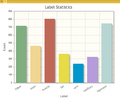 BarChartWithCategoricalData.png 576 × 486; 26 KB
 Barrott-JClinInvest2018-fig6.jpg 733 × 397; 99 KB
 Bartling-JComputAssistTomogr2005-fig.jpg 500 × 684; 169 KB
 Bartling-JComputAssistTomogr2005.pdf 1,275 × 1,650, 6 pages; 533 KB
 Base-Features-and-Modules.png 914 × 495; 119 KB
- BasePiece.zip ; 148 KB
 BaselineCBCTCMFreg.png 353 × 307; 82 KB
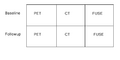
 BaselineFollowupSCANCMFreg.png 353 × 325; 85 KB
 BaselineFollowupSCANCMFreg2.png 353 × 325; 85 KB
 BaselineFollowupSCANRegisteredCMFreg.png 353 × 325; 85 KB
 BaselineFollowupSCANRegisteredCMFreg2.png 354 × 324; 86 KB
 BaselineSegmentationMaskCMFreg.png 342 × 300; 78 KB
 BasicController.png 356 × 75; 3 KB
 BasicHeartView.png 1,503 × 946; 460 KB
 Basicbraintissues gui.png 496 × 578; 42 KB
 Baumgarten-IEEE TBE2017.png 850 × 640; 244 KB
- BeatingHeart.mov ; 8.27 MB
 Before.png 919 × 478; 439 KB
 BeforeAffineRegistration.png 448 × 340; 78 KB
 Before reg.png 510 × 510; 89 KB
 Behan-FrontNeurosci2017.jpg 642 × 920; 226 KB
 Beig-SciRep2018-fig3.jpg 660 × 478; 214 KB
 Bennett-PLoSPathogens2009-fig6.png 1,035 × 610; 535 KB
 Bennett-PLoSPathogens2010.pdf 1,275 × 1,647, 10 pages; 1.69 MB
 Berman-JNeurosurg2004-fig2e.png 475 × 466; 254 KB
 Berman-JNeurosurg2004.pdf 1,218 × 1,631, 7 pages; 255 KB
 BetaProbe.jpg 2,622 × 884; 1.08 MB
 BinShrinkGoldGrayscale.png 1,600 × 1,178; 568 KB
 BinShrinkSettings.png 602 × 266; 34 KB
 Bioimagesuite slicer1.png 591 × 394; 51 KB
 Bioimagesuite slicer2.png 1,304 × 940; 94 KB
 Bioimagesuite slicer3.png 692 × 1,095; 65 KB
 Bioimagesuite slicer4.png 1,031 × 736; 63 KB
 Biolectromagnetics group UCC.jpg 1,000 × 288; 36 KB
 BlendVectorVolumes-GUI.png 488 × 499; 58 KB
 Bloblike.png 400 × 400; 43 KB
 Blocker-PLoS One 2019-fig7.jpg 700 × 469; 85 KB
 BlurImage1.png 320 × 281; 21 KB
 BlurImage2.png 320 × 281; 34 KB
 BoneTexture-BMParam.png 535 × 84; 6 KB
 BoneTexture-DisplayColormaps.png 488 × 84; 5 KB
 BoneTexture-FeatureSelection.png 645 × 171; 39 KB
 BoneTexture-FeatureValues.png 542 × 358; 44 KB
 BoneTexture-GLCMParam.png 494 × 170; 13 KB
 BoneTexture-GLRLMParam.png 493 × 222; 17 KB
 BoneTexture-Input.png 487 × 105; 6 KB
 BoneTexture-Interface.png 742 × 849; 72 KB
 BoneTexture-Parameter.png 549 × 215; 11 KB
 BoneTextureExtension-Slicer.png 1,855 × 1,056; 336 KB
 BoneTextureSerealizer-Input.png 500 × 79; 8 KB
 BoneTextureSerializer-Exportation.png 502 × 89; 9 KB
 BoneTextureSerializer-Interface.png 559 × 477; 38 KB
 Boolean-Remove.jpg 1,504 × 983; 324 KB
 BottomDeselectedIndicatorAndIconBlank.png 21 × 36; 354 bytes
 BottomDisabledIndicatorAndIconBlank.png 21 × 36; 373 bytes
 BottomSelectedCheckIndicatorAndIconBlank.png 21 × 36; 414 bytes
 BottomSelectedRadioIndicatorAndIconBlank.png 21 × 36; 390 bytes
 Boubela-Frontiers-in-Physics2014-fig4.png 2,373 × 907; 1.14 MB
 Boubela-Frontiers-in-Physics2014.pdf 1,240 × 1,624, 8 pages; 3.09 MB
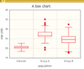 BoxChart.png 440 × 360; 22 KB
 Brain.jpg 1,433 × 1,085; 57 KB
 BrainAtlas2012.jpg 900 × 598; 274 KB
- BrainAtlasClassifier.zip ; 39.9 MB
 BrainExtractionTool-GUI.png 482 × 484; 30 KB
 BrainExtractionTool-logo.png 128 × 128; 20 KB
 BrainGrowth RegressionSequence.gif 600 × 422; 1.91 MB
 BrainH.png 200 × 175; 46 KB
 BrainH1.png 808 × 827; 538 KB
 BrainLogisticSegmentation gui.png 590 × 814; 54 KB
 BrainMRI.png 746 × 620; 106 KB
 BrainModelVolume RegressionPlot.png 2,032 × 1,654; 214 KB
 BrainSpecialist.png 202 × 247; 27 KB
 BrainStructuresSegmenter-icon.png 128 × 128; 14 KB
 BrainStructuresSegmenter gui.png 479 × 582; 52 KB
 BrainTissuesExtension-logo.png 128 × 128; 17 KB
 BrainWebImageTransformed.jpg 457 × 564; 27 KB
 Brain 3D.png 372 × 294; 133 KB
- Brain Atlas Tutorial08.ppt ; 2.99 MB
 Brain T1 orig.png 373 × 295; 10 KB
 Brain after.jpg 1,054 × 742; 153 KB
 Brain after section.jpg 645 × 692; 90 KB
 Brain before.jpg 1,054 × 742; 94 KB
 Brain section.jpg 717 × 729; 75 KB
 Brainextractiontool gui.png 482 × 484; 30 KB
 Brainlab bioimage slicer3.png 960 × 720; 72 KB
 Braintissuesmask gui.png 485 × 618; 46 KB
- Breast-data1.zip ; 62.52 MB
- Breast-data2.zip ; 56.54 MB
- BreastHeteroCADData.zip ; 62.77 MB
 Brem-AcadRadiol2007-fig1.png 500 × 429; 188 KB
 Brem-SkeletalRadiol2007-fig1.jpg 616 × 529; 151 KB
 Brem-SkeletalRadiology2007.pdf 1,240 × 1,647, 6 pages; 191 KB
 Bricault-Radiology2006-fig2c.png 713 × 613; 178 KB
 Bricault-Radiology2006.pdf 1,237 × 1,631, 8 pages; 358 KB
 Brickman-NatNeurosci2014-fig1.jpg 1,050 × 909; 167 KB
 Bucking-PLoSOne2017.jpg 704 × 253; 46 KB
 Bug.png 20 × 38; 1 KB
 BuildInstruction-CTK-build.png 857 × 640; 43 KB
 BuildInstructions-CTK-build-VTK DIR.png 408 × 234; 8 KB
 BuildInstructions-Slicer-CTK DIR.png 408 × 234; 8 KB
 BuildInstructions-Slicer-UseQt.png 857 × 640; 47 KB
 Bulbgraph.png 18 × 22; 1 KB
 Bumps conv 1.png 804 × 831; 180 KB
 Burke-JNeurosci2014-fig2.png 321 × 501; 95 KB
 Butaric-mj.png 418 × 299; 79 KB
 Butaric-slicer-mj.pdf 1,275 × 1,650, 3 pages; 428 KB
 C.gif 600 × 459; 25.09 MB
 CA-1.png 1,978 × 1,676; 143 KB
 CA-UI1.png 1,054 × 376; 28 KB
 CA-UI2.png 1,060 × 662; 54 KB
 CA-UI3.png 1,094 × 210; 24 KB
 CANARIE h small.png 1,269 × 408; 106 KB
 CANARIE h verysmall.png 423 × 136; 28 KB
 CANARIE logoonly small.png 326 × 345; 79 KB
 CAPES-logo.png 3,642 × 3,341; 1.46 MB
 CARS2014 Murakami.pdf 1,240 × 1,753, 3 pages; 504 KB
- CASE42.nrrd ; 5.05 MB
 CCSelectedTracts.png 933 × 836; 383 KB
 CC label.png 905 × 727; 105 KB
 CC tract label.png 895 × 727; 56 KB
 CDash-logo.png 270 × 270; 32 KB
 CLIAutoRun.png 1,266 × 803; 295 KB
 CMFAddAndMoveLandmarksTab.png 1,058 × 276; 35 KB
 CMFCalculateAngleBetweenLinesTab.png 1,058 × 544; 69 KB
 CMFCalculateDistanceBetweenLandmarksTab.png 1,056 × 494; 63 KB
 CMFCalculateDistanceBetweenLineLandmrksTab.png 1,056 × 448; 59 KB
 CMFChoosePlanesTab.png 978 × 168; 17 KB
 CMFClippingTab.png 980 × 474; 51 KB
 CMFDefineMiddlePointTab.png 976 × 272; 36 KB
 CMFDefineMiddlePointsTab.png 1,058 × 222; 30 KB
 CMFMangePlaneTab1.png 978 × 390; 45 KB
 CMFMangePlaneTab2.png 978 × 352; 33 KB
 CMFPropagationTab.png 1,090 × 202; 24 KB
 CMFROITab.png 1,088 × 440; 58 KB
 CMFResultsTab.png 980 × 350; 25 KB
 CMFSaveTab.png 982 × 200; 24 KB
 CMFSceneTab.png 982 × 402; 29 KB
 CMFVisibilityOfModelsAndLandmarksTab.png 1,058 × 306; 23 KB
- CMFregData.zip ; 315.23 MB
 CMRTK-logo.png 128 × 128; 27 KB
 CMRToolkit.png 185 × 185; 49 KB
 CMRToolkitWizard Panel1.png 541 × 770; 33 KB
 CMRToolkitWizard Panel2.png 538 × 768; 59 KB
 CMRToolkitWizard Panel3.png 536 × 768; 32 KB
 CMRToolkit GraphCut.png 1,392 × 934; 338 KB
 CMRToolkit Threshold Model.gif 450 × 209; 304 KB
 CMRToolkit Threshold Model.png 1,392 × 934; 232 KB
- CMRToolkit Threshold Model Tutorial.pptx ; 2.02 MB
 CMRToolkit Tutorial Image.png 599 × 487; 99 KB
 CMRToolkit UofU logo.jpg 592 × 129; 51 KB
 CMakeLogoMediumResolution.png 518 × 518; 48 KB
 CMakePerformance.png 1,411 × 857; 49 KB
 CMakePerformance4144.png 1,833 × 1,066; 64 KB
 CMakePerformance415Nightly.png 1,828 × 1,060; 68 KB
 CNPq-logo.png 3,999 × 1,200; 158 KB
 CNPqOrig-logo.png 880 × 265; 25 KB
 CRTC logo.jpg 75 × 76; 8 KB
 CSAIL-logo.png 490 × 408; 72 KB
 CSIM-logo.png 1,097 × 1,029; 70 KB
- CT-angio-04-2009.zip ; 111.64 MB
- CT-chest.nrrd ; 40.23 MB
 CT-chest screenshot.png 779 × 684; 256 KB
- CTA-cardio.nrrd ; 61.08 MB
 CTHandBone BeforeAfter.png 692 × 332; 154 KB
 CTK-Logo.png 630 × 630; 194 KB
 CTKApplauncher Logo.png 200 × 200; 19 KB
 CTSC BC PETCTfuse.png 400 × 200; 5 KB
- CT liver segmentation case.tgz ; 62.91 MB
 Caan-MIA2006-fig8.png 1,103 × 455; 139 KB
 Caan-MIA2006.pdf 1,240 × 1,653, 9 pages; 570 KB
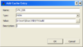
 CacheSettings.png 706 × 497; 20 KB
 Cadotte-pone2015-fig6.jpg 683 × 959; 94 KB
 Calculator-Screenshot.jpg 1,457 × 863; 218 KB
 Calculator.png 128 × 128; 31 KB
 Calculus.png 150 × 128; 22 KB
 Calculus module ui.png 499 × 550; 17 KB
 Calculus module ui1.png 498 × 638; 17 KB
 Camerapath module.png 434 × 723; 81 KB
 Canine-heart1.png 1,248 × 1,109; 266 KB
 Caption.jpg 848 × 642; 98 KB
 Caption.png 446 × 240; 19 KB
 Caption1.PNG 852 × 650; 367 KB
 Caption1.png 852 × 650; 367 KB
 Caption2.PNG 379 × 360; 13 KB
 Caption2.jpg 379 × 371; 33 KB
 Caption2.png 379 × 371; 33 KB
 Caption3.PNG 855 × 294; 22 KB
 Caption3.png 301 × 236; 11 KB
 Caption4.jpg 379 × 371; 33 KB
 Caption5.jpg 377 × 373; 33 KB
 Caption6.jpg 845 × 633; 107 KB
 Captura2.jpg 379 × 371; 33 KB
 Capture1.png 301 × 236; 11 KB
 Capture2.png 387 × 275; 15 KB
 Carasa-PlosOne2017-fig2.jpg 800 × 764; 98 KB
 CardiacAgatstonMeasuresModuleScreenshot.jpg 510 × 359; 51 KB
 CardiacAgatstonModuleIamge.png 128 × 128; 19 KB
- CardiacCT.seq.nrrd ; 12.49 MB
 CardiacCtReplay.gif 481 × 325; 389 KB
- Cardiac ECGg CT.tgz ; 12.81 MB
- Cardiac MRI Toolkit Tutorial Data.zip ; 10.14 MB
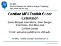 Cardiac MRI Toolkit Tutorial Summer2013.pdf 1,500 × 1,125, 41 pages; 2.4 MB
- Cardiac MRI Toolkit Tutorial Summer2013.pptx ; 156.19 MB
 Cardiac mri registration module.jpg 1,503 × 1,029; 735 KB
 Cardinale-SEEG Scarabin2012.png 378 × 246; 156 KB
 Cardioseg+volume.png 1,509 × 1,128; 478 KB
 Carlson-mj.pdf 1,239 × 1,629, 12 pages; 164 KB
 Carlson-mj.png 430 × 600; 201 KB
 CarmaAxialDilate1.png 1,259 × 695; 58 KB
 CarmaAxialDilate2.png 1,259 × 695; 62 KB
- Carma Boolean Remove Tutorial.pptx ; 1.57 MB
- Carma PVAntrumCutFilter Tutorial.pptx ; 1.61 MB
- Carma Registration Tutorial.pptx ; 2.87 MB
 Carma UofU logo.jpg 1,035 × 542; 204 KB
 Carma afib auto scar.png 1,443 × 900; 640 KB
 Carma afib inhomogeneity correction.png 1,443 × 900; 672 KB
 CarotidSegmentation.png 1,302 × 744; 232 KB
 CarotidSegmentationROI.png 1,300 × 745; 178 KB
 CarreraSliceBlogEntry.png 1,056 × 816; 324 KB
 CarreraSliceEffect.png 21 × 21; 555 bytes
 CartilageMRI-LUTs-2012-02-18.png 192 × 56; 9 KB
 CartilegeMIdGEMRIC15T.png 52 × 21; 6 KB
 CartilegeMIdGEMRIC3T.png 52 × 21; 6 KB
 CartilegeMRIdGEMRIC15T.png 52 × 21; 6 KB
 Caruana-FrontHumNeurosci2014-fig2.png 777 × 299; 88 KB
 Caruana-FrontHumNeurosci2014.pdf 1,240 × 1,625, 9 pages; 680 KB
 Caruana-PLoSOne2014-fig2.png 1,220 × 961; 907 KB
- Case1 DCE.tgz ; 51.25 MB
 Case1 input.png 1,433 × 1,085; 96 KB
 Case1 output.png 1,433 × 1,085; 155 KB
 Case2 input.png 1,433 × 1,085; 106 KB
 Case2 output.png 1,433 × 1,085; 188 KB
 CaseComparisonJHU.png 1,103 × 863; 512 KB
 Caskey-PLoSONE2011-fig6.png 897 × 717; 239 KB
 Caskey-journal.pone.0027372-fig3.png 1,174 × 439; 321 KB
 Catalyst logo final150.png 150 × 106; 16 KB
 Cavallari-PLosOne2014-fig1.png 320 × 241; 66 KB
 Cc.PNG 1,070 × 661; 312 KB
 Cc 01.png 754 × 508; 109 KB
 Cc stoch.png 753 × 509; 135 KB
 CellMultiChannel.png 1,914 × 1,149; 1.53 MB
 Centerlines classdiagram.png 586 × 1,056; 157 KB
 Centerlines pipeline.png 554 × 512; 21 KB
 Centerlinesgui.png 2,421 × 1,618; 636 KB
 Chadda-PLoSOne2016-fig3.jpg 733 × 231; 44 KB
 Chae-Brachytherapy2018-fig3.jpg 800 × 844; 110 KB
 Chalupa-Symmetry2018-fig1.png 314 × 332; 73 KB
 Chalupa-Symmetry2018.pdf 1,240 × 1,753, 9 pages; 1.26 MB
 ChangeIsland.png 25 × 25; 359 bytes
 ChangeIslandsEffectIcon.png 35 × 34; 4 KB
 ChangeLabel.png 25 × 25; 278 bytes
- ChangeTracker.zip ; 27.79 MB
- ChangeTracker2011.zip ; 11.04 MB
 ChangeTracker Analysis-3.4.png 1,280 × 775; 161 KB
 ChangeTracker Analysis.png 1,280 × 723; 296 KB
 ChangeTracker Step1.png 396 × 380; 5 KB
 ChangeTracker Step2 1.png 384 × 323; 4 KB
 ChangeTracker Step2 2.png 430 × 246; 89 KB
 ChangeTracker Step3.png 1,280 × 723; 182 KB
 ChangeTracker Step4.png 396 × 304; 4 KB
 ChangeTracker Step4 3-6.png 396 × 344; 6 KB
 ChangeTracker logo.png 128 × 128; 21 KB
 ChangeTracker snapshot.png 375 × 380; 12 KB
 ChangeTracker tutorial data.png 371 × 317; 5 KB
 Check.svg 16 × 16; 1 KB
 CheckAll.png 35 × 35; 910 bytes
 CheckBasalAndIctalMask.png 1,366 × 731; 134 KB
 CheckIndicatorOn.png 11 × 11; 264 bytes
 CheckModified.png 35 × 35; 894 bytes
 CheckModifiedData.png 35 × 35; 848 bytes
 CheckerBoard1.png 1,400 × 997; 514 KB
 CheckerBoard2.png 1,400 × 997; 527 KB
 Checkmark.png 20 × 15; 133 bytes
 Chen-mj.png 644 × 237; 91 KB
 ChenW-BMCMedInformDecisMak2009-fig1.jpg 600 × 622; 175 KB
 ChenW-BMCMedInformDecisMak2009.pdf 1,270 × 1,650, 14 pages; 2.38 MB
 Cherbuin-BrainBehav2011-fig1.jpg 233 × 480; 68 KB
 Cherbuin-BrainBehav2011-fig1.png 560 × 273; 118 KB
 Cherbuin-PLoS-ONE2009-fig1.png 951 × 249; 118 KB
 Cherbuin-PLoS-ONE2009.pdf 1,275 × 1,647, 10 pages; 672 KB
 Chest Imaging Platform.png 1,041 × 1,043; 965 KB
 Chick.png 178 × 233; 31 KB
 Child-i.png 100 × 100; 17 KB
 Chiu-FluidsBarriersCNS2012-fig2.png 2,362 × 2,049; 2.33 MB
 ChooseBrushSize.png 21 × 21; 543 bytes
 ChooseColor.png 21 × 21; 264 bytes
 Chow-NatNeuroci2017-fig1.png 817 × 422; 224 KB
 Christopher-CardSoc2011-fig6.png 631 × 477; 70 KB
 Christopher-CardSoc2011.pdf 1,275 × 1,650, 2 pages; 45 KB
 ChuRouen.png 1,646 × 1,243; 1.27 MB
 CineBasicController.png 356 × 75; 3 KB
 CineDisplay5.png 408 × 330; 8 KB
 CineDisplayDesigns.png 1,728 × 390; 32 KB
 CineDisplayDesigns1-4.png 455 × 1,518; 47 KB
 CirclesDetectionHoughTransform-result.png 3,012 × 1,830; 565 KB
 CiscoAnyConnectInstallOptions.png 620 × 440; 73 KB
 Civility.png 1,213 × 715; 318 KB
 Class parser state coll graph.png 635 × 1,291; 24 KB
 ClassvtkMRMLNode inherit graph.png 843 × 1,287; 175 KB
 Clauss-SocCognAffectNeurosci2014-fig1.jpg 800 × 873; 125 KB
 CleaverCIBC.png 495 × 245; 165 KB
 CleaverExtension.png 128 × 128; 30 KB
- CleaverSampleData.zip ; 1.14 MB
 Cli icon.png 960 × 720; 63 KB
 Climb.png 1,020 × 984; 794 KB
 ClipModel-with-widget.png 584 × 464; 147 KB
 Clipmodel.png 1,439 × 870; 271 KB
 Clipping.png 1,318 × 964; 413 KB
 Close.png 21 × 21; 333 bytes
 Clouard-FrontPsychol2014-fig4.jpg 746 × 511; 109 KB
 Clouard-FrontPsychol2014.pdf 1,240 × 1,625, 12 pages; 2.66 MB
 ClusterProminence.eps 339 × 454; 34 KB
 ClusterShade.eps 338 × 454; 29 KB
 CoarseSlicer2EditorWorkflow.png 800 × 650; 59 KB
 Cobb-BiomedicalComputationReview2007-fig.png 1,434 × 504; 245 KB
 Cobb-BiomedicalComputationReview2007.pdf 1,275 × 1,650, 9 pages; 616 KB
 Codelite-settings-1.png 999 × 586; 66 KB
 Codelite-settings-2.png 999 × 586; 54 KB
- Collidingfronts.swf ; 38.08 MB
 Colombo-PlosOne2015-fig2.jpg 764 × 558; 118 KB
 Color-image-reslice-2008-06-11.png 1,238 × 930; 405 KB
 ColorBar.jpg 172 × 426; 9 KB
 CommandLineModule.png 518 × 505; 109 KB
 Comment.png 20 × 38; 435 bytes
 CompareDetections.png 1,364 × 727; 176 KB
- CompareViewExample.mov ; 13.14 MB
 CompareView snapshot.png 900 × 581; 360 KB
 CompareVolumeStatistics.png 1,007 × 259; 8 KB
 CompareVolumes-1-volume.png 1,076 × 716; 667 KB
 CompareVolumes-4.4.png 1,662 × 1,205; 765 KB
 Comparison T1 Mapping ALL.png 2,318 × 1,242; 78 KB
 CompleQueryAbdominalRes.jpg 618 × 585; 142 KB
 ComplexQueryAPI.jpg 636 × 609; 150 KB
 ComplexQueryAbdominal.jpg 628 × 596; 157 KB
 ComplexQueryAbdominalWithImage.jpg 410 × 357; 83 KB
 ComplexQueryWithImage.jpg 525 × 565; 170 KB
 ComplexVis.png 841 × 764; 689 KB
 ComputationAverage-1.png 892 × 754; 78 KB
 ComputationAverage-computation.png 878 × 236; 42 KB
 ComputationAverage-preview.png 876 × 472; 62 KB
 ComputeBMFeatureMaps-Interface.png 566 × 207; 22 KB
 ComputeBMFeatures-Interface.png 564 × 375; 33 KB
 ComputeBasanAndIctalMask.png 1,366 × 728; 61 KB
 ComputeGLCMFeatureMaps-Interface.png 506 × 288; 30 KB
 ComputeGLCMFeatures-Interface.png 507 × 522; 38 KB
 ComputeGLRLMFeatureMaps-Interface.png 507 × 336; 33 KB
 ComputeGLRLMFeatures-Interface.png 505 × 623; 56 KB
 ComputeSUVBodyWeightUI.png 576 × 385; 39 KB
 CondileMask.eps 683 × 902; 30 KB
 CongealingCLIbottom.png 459 × 526; 21 KB
 CongealingCLIdefault.png 494 × 608; 27 KB
 CongealingCLItop.png 466 × 703; 27 KB
 ConnectAB2.png 738 × 501; 45 KB
 ConnectedThreshold.jpg 1,503 × 925; 570 KB
 ConnectivityMap.png 1,409 × 995; 416 KB
 Connectmenu.png 359 × 134; 2 KB
 ContextMenu.png 469 × 323; 108 KB
 ContinuousRedGreenBlue.png 35 × 24; 749 bytes
- ContinuousRedGreenBlue.tif 35 × 24; 1 KB
 Contour2d.png 378 × 323; 67 KB
 Contour3dVolume.png 378 × 323; 79 KB
 ContourRepresentations 201507.png 767 × 845; 122 KB
 Contributing3DSlicerExtension.png 931 × 698; 156 KB
- ControlledVocabulary.xls ; 159 KB
 Conventional-Widescreen-10-2011.png 2,334 × 1,415; 1.8 MB
 ConvertGuide2ROI.png 21 × 21; 344 bytes
 ConvertROI2Label.png 21 × 21; 340 bytes
 Converters-2013-02-21.png 421 × 88; 18 KB
 Coordinate sytems.png 981 × 407; 197 KB
 CopyPasteSlice.png 1,600 × 1,152; 361 KB
 CopyROI.png 21 × 21; 345 bytes
 CoreModules-2013-02-21.png 226 × 252; 25 KB
 Cornella-AJOG2003-fig2.png 690 × 679; 604 KB
 Cornella-AJOG2003.pdf 1,218 × 1,631, 6 pages; 2.51 MB
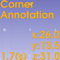
 CornerAnnotation1.png 2,154 × 1,448; 263 KB
 CornerAnnotation2.png 2,258 × 1,412; 396 KB
 CornerAnnotationIcon.png 128 × 128; 23 KB
 CornerAnnotationModuleSS.png 2,878 × 1,670; 724 KB
 Correlation.eps 336 × 454; 32 KB
 Corticalthickness.png 320 × 198; 81 KB
 Cover-HeartRhythm2008.png 612 × 630; 513 KB
 Covington-AnnuORNLBiomedSciEngCentConf2010-fig4.png 571 × 663; 266 KB
 Cramer-PloSOne2015-fig3.jpg 632 × 560; 69 KB
 CreateFiducialClickHere.png 1,600 × 1,200; 532 KB


