Main Page/SlicerCommunity/2020
Go to 2022 :: 2021 :: 2020 :: 2019 :: 2018 :: 2017 :: 2016 :: 2015 :: 2014-2011 :: 2010-2000
The community that relies on 3D Slicer is large and active: (numbers below updated on December 1st, 2023)
- 1,467,466+ downloads in the last 11 years (269,677 in 2023, 206,541 in 2022)
- over 17.900+ literature search results on Google Scholar
- 2,147+ papers on PubMed citing the Slicer platform paper
- Fedorov A., Beichel R., Kalpathy-Cramer J., Finet J., Fillion-Robin J-C., Pujol S., Bauer C., Jennings D., Fennessy F.M., Sonka M., Buatti J., Aylward S.R., Miller J.V., Pieper S., Kikinis R. 3D Slicer as an Image Computing Platform for the Quantitative Imaging Network. Magnetic Resonance Imaging. 2012 Nov;30(9):1323-41. PMID: 22770690. PMCID: PMC3466397.
- 39 events in open source hackathon series continuously running since 2005 with 3260 total participants
- Slicer Forum with +8,138 subscribers has approximately 275 posts every week
The following is a sample of the research performed using 3D Slicer outside of the group that develops it. in 2020
We monitor PubMed and related databases to update these lists, but if you know of other research related to the Slicer community that should be included here please email: marianna (at) bwh.harvard.edu.
Contents
- 1 2020
- 1.1 Temporomandibular Joint Damage in K/BxN Arthritic Mice
- 1.2 Visualization of Mucosal Field in HPV Positive and Negative Oropharyngeal Squamous Cell Carcinomas: Combined Genomic and Radiology Based 3D Model
- 1.3 Manual and Semiautomatic Segmentation of Bone Sarcomas on MRI Have High similarity
- 1.4 Measurement Error and Reliability of Three Available 3D Superimposition Methods in Growing Patients
- 1.5 Prostate Multiparametric Magnetic Resonance Imaging Features Following Partial Gland Cryoablation
2020
Temporomandibular Joint Damage in K/BxN Arthritic Mice
|
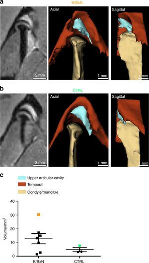
Publication: Int J Oral Sci. 2020 Feb 6;12(1):5. PMID: 32024813 | PDF Authors: Kuchler-Bopp S, Mariotte A, Strub M, Po C, De Cauwer A, Schulz G, Van Bellinghen X, Fioretti F, Clauss F, Georgel P, Benkirane-Jessel N, Bornert F. Institution: INSERM (French National Institute of Health and Medical Research), UMR 1260, Regenerative NanoMedicine (RNM), FMTS, Strasbourg, France. Abstract: Rheumatoid arthritis (RA) is an autoimmune disease affecting 1% of the world population and is characterized by chronic inflammation of the joints sometimes accompanied by extra-articular manifestations. K/BxN mice, originally described in 1996 as a model of polyarthritis, exhibit knee joint alterations. The aim of this study was to describe temporomandibular joint (TMJ) inflammation and damage in these mice. We used relevant imaging modalities, such as micro-magnetic resonance imaging (μMRI) and micro-computed tomography (μCT), as well as histology and immunofluorescence techniques to detect TMJ alterations in this mouse model. Histology and immunofluorescence for Col-I, Col-II, and aggrecan showed cartilage damage in the TMJ of K/BxN animals, which was also evidenced by μCT but was less pronounced than that seen in the knee joints. μMRI observations suggested an increased volume of the upper articular cavity, an indicator of an inflammatory process. Fibroblast-like synoviocytes (FLSs) isolated from the TMJ of K/BxN mice secreted inflammatory cytokines (IL-6 and IL-1β) and expressed degradative mediators such as matrix metalloproteinases (MMPs). K/BxN mice represent an attractive model for describing and investigating spontaneous damage to the TMJ, a painful disorder in humans with an etiology that is still poorly understood. The volume estimation and 3D reconstructions were obtained with 3D Slicer. |
 Comparison of synovial fluid volume of K/BxN and control TMJs. Magnetic resonance imaging (MRI) and 3D reconstructions of the TMJ of a K/BxN mouse (a) and a control mouse (b). c Volume measurement of the upper articular cavity of seven K/BxN mice and three control mice. The orange square represents the K/BxN mouse shown in a, and the green circle represents the control mouse shown in b. |
Visualization of Mucosal Field in HPV Positive and Negative Oropharyngeal Squamous Cell Carcinomas: Combined Genomic and Radiology Based 3D Model
|
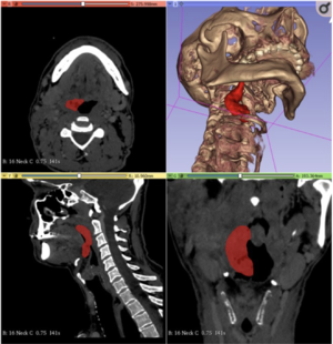
Publication: Sci Rep. 2020 Jan 8;10(1):40. PMID: 31913295 | PDF Authors: Orosz E, Gombos K, Petrevszky N, Csonka D, Haber I, Kaszas B, Toth A, Molnar K, Kalacs K, Piski Z, Gerlinger I, Burian A, Bellyei S, Szanyi I. Institution: University of Pécs, Medical School, Department of Otorhinolaryngology, Pécs, Hungary. Abstract: The aim of this study was to visualize the tumor propagation and surrounding mucosal field in radiography-based 3D model for advanced stage HNSCC and combine it with HPV genotyping and miRNA expression characterization of the visualized area. 25 patients with T1-3 clinical stage HNSCC were enrolled in mapping biopsy sampling. Biopsy samples were evaluated for HPV positivity and miR-21-5p, miR-143, miR-155, miR-221-5p expression in Digital Droplet PCR system. Significant miRNA expression differences of HPV positive tumor tissue biopsies were found for miR-21-5p, miR-143 and miR-221-5p compared to the HPV negative tumor biopsy series. Peritumoral mucosa showed patchy pattern alterations of miR-21-5p and miR-155 in HPV positive cases, while gradual change of miR-21-5p and miR-221-5p was seen in HPV negative tumors. In our study we found differences of the miRNA expression patterns among the HPV positive and negative tumorous tissues as well as the surrounding mucosal fields. The CT based 3D models of the cancer field and surrounding mucosal surface can be utilized to improve proper preoperative planning. Complex evaluation of HNSCC tissue organization field can elucidate the clinical and molecular differentiation of HPV positive and negative cases, and enhance effective organ saving therapeutic strategies. |
] |
Manual and Semiautomatic Segmentation of Bone Sarcomas on MRI Have High similarity
|
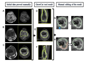
Publication: Braz J Med Biol Res. 2020 Jan 31;53(2):e8962. PMID: 32022102 | PDF Authors: Dionísio FCF, Oliveira LS, Hernandes MA, Engel EE, Rangayyan RM, Azevedo-Marques PM, Nogueira-Barbosa MH. Institution: Departamento de Imagens Médicas, Hematologia e Oncologia Clínica, Faculdade de Medicina de Ribeirão Preto, Universidade de São Paulo, Ribeirão Preto, SP, Brasil. Abstract: The aims of this study were to evaluate the intra- and interobserver reproducibility of manual segmentation of bone sarcomas in magnetic resonance imaging (MRI) studies and to compare manual and semiautomatic segmentation methods. This retrospective study included twelve osteosarcoma and eight Ewing sarcoma MRI studies performed prior to any therapeutic intervention. All cases were histopathologically confirmed. Three radiologists used 3D Slicer software to perform manual segmentation of bone sarcomas in a blinded and independent manner. One radiologist segmented manually and also performed semiautomatic segmentation with the GrowCut tool. Segmentation exercises were timed for comparison. The dice similarity coefficient (DSC) and Hausdorff distance (HD) were used to evaluate similarity between the segmentation results and further statistical analyses were performed to compare DSC, HD, and volumetric results. Manual segmentation was reproducible with intraobserver DSC varying from 0.83 to 0.97 and HD from 3.37 to 28.73 mm. Interobserver DSC of manual segmentation showed variation from 0.73 to 0.97 and HD from 3.93 to 33.40 mm. Semiautomatic segmentation compared to manual segmentation resulted in DSCs of 0.71-0.96 and HDs of 5.38-31.54 mm. Semiautomatic segmentation required significantly less time compared to manual segmentation (P value ≤0.05). Among all situations compared, tumor volumetry did not show significant statistical differences (P value >0.05). We found excellent intra- and interobserver agreement for manual segmentation of osteosarcoma and Ewing sarcoma. There was high similarity between manual and semiautomatic segmentation, with a significant reduction of segmentation time using the semiautomatic method. |
 3D Slicer’s GrowCut semiautomatic segmentation steps of a femoral osteosarcoma magnetic resonance imaging case. In the first column (A, B, and C), the T1WI sequence images are on the left and the T1WI FS GD sequence images are on the right. In the second (D, E, and F) and third columns (G and H), there are only T1WI FS GD sequence images. A, Yellow rectangular area of background tissue beyond the inferior margin of tumor. B and C, Area of interest from the tumor tissue within green mark and background tissue within yellow mark in the extremities of the tumor (B) and in the middle portion of the tumor on the longitudinal axis (C). D, E, and F, Segmented volume of interest within green mark after GrowCut tool processing in coronal (D), axial (E), and sagittal (F) planes. G and H, Manual editing of GrowCut tool excluding areas from the volume of interest (within green mark) that do not contain tumor tissue, as indicated with the blue arrow in (G), and including areas with tumor tissue in the volume of interest, as shown in area marked in red (H). ] |
Measurement Error and Reliability of Three Available 3D Superimposition Methods in Growing Patients
|
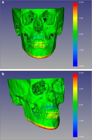
Publication: Head Face Med. 2020 Jan 27;16(1):1. PMID: 31987041 | PDF Authors: Ponce-Garcia C, Ruellas ACO, Cevidanes LHS, Flores-Mir C, Carey JP, Lagravere-Vich M. Institution: Division of Orthodontics, School of Dentistry, University of Alberta, Edmonton, Canada. Abstract: Cone-Beam Computed Tomography (CBCT) images can be superimposed, allowing three-dimensional (3D) evaluation of craniofacial growth/treatment effects. Limitations of 3D superimposition techniques are related to imaging quality, software/hardware performance, reference areas chosen, and landmark points/volumes identification errors. The aims of this research are to determine/compare the intra-rater reliability generated by three 3D superimposition methods using CBCT images, and compare the changes observed in treated cases by these methods. METHODS: Thirty-six growing individuals (11-14 years old) were selected from patients that received orthodontic treatment. Before and after treatment (average 24 months apart) CBCTs were analyzed using three superimposition methods. The superimposed scans with the two voxel-based methods were used to construct surface models and quantify differences using 3D SlicerCMF software, while distances in the landmark-derived method were calculated using Excel. 3D linear measurements of the models superimposed with each method were then compared. RESULTS: Repeated measurements with each method separately presented good to excellent intraclass correlation coefficient (ICC ≥ 0.825). ICC values were the lowest when comparing the landmark-based method and both voxel-based methods. Moderate to excellent agreement was observed when comparing the voxel-based methods against each other. The landmark-based method generated the highest measurement error. CONCLUSIONS: Findings indicate good to excellent intra-examiner reliability of the three 3D superimposition methods when assessed individually. However, when assessing reliability among the three methods, ICC demonstrated less powerful agreement. The measurements with two of the three methods (CMFreg/3D Slicer and Dolphin) showed similar mean differences; however, the accuracy of the results could not be determined. |
 Color-coded map with CMFreg/3D Slicer method for visualization purposes only, not quantitative assessment. Frontal (4a) and 45 degrees (4b) views of the 3D color-coded maps showing the change in mm. ] |
Prostate Multiparametric Magnetic Resonance Imaging Features Following Partial Gland Cryoablation
|
Publication: Urology. 2020 Jan 15. PMID: 31954170 Authors: Al Awamlh BAH, Margolis DJ, Gross MD, Natarajan S, Priester A, Hectors S, Ma X, Mosquera JM, Liao J, Hu JC. Institution: Division of Orthodontics, School of Dentistry, University of Alberta, Edmonton, Canada. Abstract: OBJECTIVES: To assess the qualitative and quantitative changes on prostate multiparametric magnetic resonance imaging (mpMRI) following partial gland ablation (PGA) with cryotherapy and correlate with histopathology. METHODS: We used 3D Slicer to generate prostate models and segment ipsilateral (treated) and contralateral peripheral and transition zones in ten men who underwent MRI/transrectal ultrasound fusion-guided PGA during 2017-2018. Pre and post-PGA volumes of prostate segments were compared. Post-PGA mpMRI were categorized according to PI-RADS v2 and treatment response on mpMRI was assessed in a manner similar to the radiology evaluation framework following liver lesion ablation. RESULTS: Median volume of ipsilateral peripheral and transition zones decreased from 10.9 mL and 13.0 mL to 7.2 mL and 10.8 mL (p=0.005), respectively. Median volume of contralateral peripheral and transition zones also decreased from 12.1 mL and 12.5 mL to 9.9 mL to 10.4 mL (p=0.005), respectively. Five men had clinically significant disease (Grade group ≥2) on post-PGA biopsy (three within treatment field and two outside). Of the men with clinically significant prostate cancer, mpMRI revealed PI-RADS 3 lesions in two. However, the treatment response framework did not detect residual disease. CONCLUSION: PGA results in asymmetric and significant reductions in prostate volume. Our results highlight the need for a separate assessment framework to enable standardization of the interpretation and reporting of post-PGA surveillance mpMRI. Moreover, our findings have significant implications for MRI-targeted surveillance biopsy following PGA with cryotherapy.
|
