File:Slicer-48-Release blogpost-350x329.gif
Slicer-48-Release_blogpost-350x329.gif (350 × 329 pixels, file size: 1.43 MB, MIME type: image/gif, looped, 31 frames)
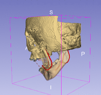
3-dimensional reconstruction of craniofacial hard tissue (bone, in solid/semitransparent yellow) and soft tissue (vessels in solid red, gingiva in semitransparent pink) structures, rendered altogether with orthodontic appliances (brackets in solid blue) obtained from Cone Beam Computed Tomography. This data was generated as surgical scenario for Bisagittal Split Osteotomy (BSSO) training. The models in this figure represent the pre-surgical state of the patient, and Slicer 4.8 will help the trainee practicing all the different steps of BSSO, in which (1) the gingiva is cut to expose the bone, (2) the bone is cut bilaterally in the mandible into distal and proximal segments, (3) the distal segment of the mandible is moved forward and finally (4) stabilized in its new position in the proximal segment with fixation screws. BSSO is performed to return patient skeletal structures into their proper alignment in cases of mandibular deficiency. This work was supported by the National Institute of Health (NIH) National Institute for Dental and Craniofacial Research (NIDCR) grant R43DE027595 (High-Fidelity Virtual Reality Trainer for Orthognathic Surgery).
File history
Click on a date/time to view the file as it appeared at that time.
| Date/Time | Thumbnail | Dimensions | User | Comment | |
|---|---|---|---|---|---|
| current | 20:05, 26 October 2017 | | 350 × 329 (1.43 MB) | JChris.FillionR (talk | contribs) | 3-dimensional reconstruction of craniofacial hard tissue (bone, in solid/semitransparent yellow) and soft tissue (vessels in solid red, gingiva in semitransparent pink) structures, rendered altogether with orthodontic appliances (brackets in solid blue... |
- You cannot overwrite this file.
File usage
There are no pages that use this file.