EMSegmenter-Tasks:Non-Human-Primate
From Slicer Wiki
Home < EMSegmenter-Tasks:Non-Human-Primate
Return to EMSegmenter Task Overview Page
Description
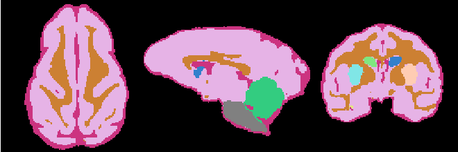
Non Human Primate pipeline ...TODO
The pipeline consist of the following steps:
- Step 1: Perform image inhomogeneity correction of the MRI scan via N4ITKBiasFieldCorrection (Tustison et al 2010)
- Step 2: Register the atlas to the MRI scan via BRAINSFit (Johnson et al 2007)
- Step 3: Compute the intensity distributions for each structure
Compute intensity distribution (mean and variance) for each label by automatically sampling from the MR scan. The sampling for a specific label is constrained to the region that consists of voxels with high probability (top 95%) of being assigned to the label according to the aligned atlas.
- Step 4: Automatically segment the MRI scan into the structures of interest using EM Algorithm (Pohl et al 2007)
Anatomical Tree
- Root
- Head
- intracranial cavity (ICC)
- White matter + Gray matter + CSF class (WGC)
- grey matter (GM)
- left Subcortical Gray Matter (lSGM)
- left caudate (lCaudate)
- left putamen (lPutamen)
- left hippocampus (lHippocampus)
- right Subcortical Gray Matter (rSGM)
- right caudate (rCaudate)
- right putamen (rPutamen)
- right hippocampus (rHippocampus)
- Cortical Gray Matter (CGM)
- left Subcortical Gray Matter (lSGM)
- white matter (WM)
- cerebrospinal fluid (CSF)
- grey matter (GM)
- Brainstem
- Cerebellum
- White matter + Gray matter + CSF class (WGC)
- background (BG)
- intracranial cavity (ICC)
- Head
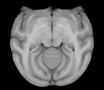
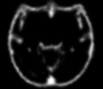
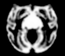
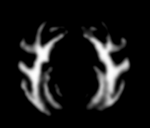
Atlas
Image Dimension = 256 x 256 x 252
Image Spacing = 0.4688 x 0.4688 x 0.50002
|
|
|
|
| Template (T1) | CSF | GM | WM |
Result
Collaborators
Andriy Fedorov (BWH)
Citations
- Tustison NJ, Avants BB, Cook PA, Zheng Y, Egan A, Yushkevich PA, Gee JC N4ITK: Improved N3 Bias Correction, IEEE Trans Med Imag, 2010
- Pohl K, Bouix S, Nakamura M, Rohlfing T, McCarley R, Kikinis R, Grimson W, Shenton M, Wells W. A Hierarchical Algorithm for MR Brain Image Parcellation. IEEE Transactions on Medical Imaging. 2007 Sept;26(9):1201-1212.
- S. Warfield, J. Rexilius, P. Huppi, T. Inder, E. Miller, W. Wells, G. Zientara, F. Jolesz, and R. Kikinis, “A binary entropy measure to assess nonrigid registration algorithms,” in MICCAI, LNCS, pp. 266–274, Springer, October 2001.
- Johnson H.J., Harris G., Williams K. BRAINSFit: Mutual Information Registrations of Whole-Brain 3D Images, Using the Insight Toolkit, The Insight Journal, July 2007

