Difference between revisions of "EMSegmenter-Tasks:MRI-Human-Brain-Parcellation"
From Slicer Wiki
Belhachemi (talk | contribs) (→Result) |
Belhachemi (talk | contribs) |
||
| Line 1: | Line 1: | ||
| − | + | =Description= | |
MRI Human Head pipeline for a finer-grained parcellation (Priya please provide description and proper citation) | MRI Human Head pipeline for a finer-grained parcellation (Priya please provide description and proper citation) | ||
| − | + | =Anatomical Tree= | |
The anatomical tree represents the structures to be segmented. Node labels displayed below contain a human readable structure name and in parentheses the internally used structure name. | The anatomical tree represents the structures to be segmented. Node labels displayed below contain a human readable structure name and in parentheses the internally used structure name. | ||
| Line 34: | Line 34: | ||
** cerebrospinal fluid (CSF) | ** cerebrospinal fluid (CSF) | ||
| − | + | =Atlas= | |
The atlas hes been created by Padmapriya Srinivasan and Sylvain Bouix from (PNL-BWH) | The atlas hes been created by Padmapriya Srinivasan and Sylvain Bouix from (PNL-BWH) | ||
| Line 42: | Line 42: | ||
'''TODO, some details about atlas''' | '''TODO, some details about atlas''' | ||
| − | + | =Pre-Processing= | |
* 1 step: image inhomogeneity correction [http://www.slicer.org/slicerWiki/index.php/Modules:N4ITKBiasFieldCorrection-Documentation-3.6 N4ITKBiasFieldCorrection] | * 1 step: image inhomogeneity correction [http://www.slicer.org/slicerWiki/index.php/Modules:N4ITKBiasFieldCorrection-Documentation-3.6 N4ITKBiasFieldCorrection] | ||
* 2 step: atlas-to-target registration [http://www.slicer.org/slicerWiki/index.php/Modules:BRAINSFit BRAINSFit] | * 2 step: atlas-to-target registration [http://www.slicer.org/slicerWiki/index.php/Modules:BRAINSFit BRAINSFit] | ||
| Line 51: | Line 51: | ||
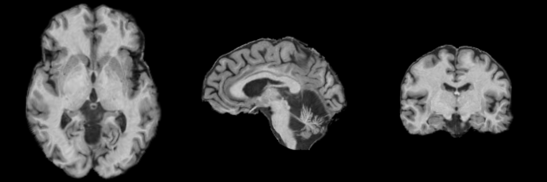
[[Image:MRI-Human-Brain-Parcellation_3x360x360.png]] | [[Image:MRI-Human-Brain-Parcellation_3x360x360.png]] | ||
| − | + | =Collaborators= | |
Padmapriya Srinivasan and Sylvain Bouix (PNL-BWH) | Padmapriya Srinivasan and Sylvain Bouix (PNL-BWH) | ||
| + | |||
| + | =Citations= | ||
| + | * Tustison NJ, Avants BB, Cook PA, Zheng Y, Egan A, Yushkevich PA, Gee JC N4ITK: Improved N3 Bias Correction, IEEE Trans Med Imag, 2010 | ||
| + | * Pohl K, Bouix S, Nakamura M, Rohlfing T, McCarley R, Kikinis R, Grimson W, Shenton M, Wells W. [http://www.slicer.org/pages/Special:PubDB_View?dspaceid=608 A Hierarchical Algorithm for MR Brain Image Parcellation.] IEEE Transactions on Medical Imaging. 2007 Sept;26(9):1201-1212. | ||
| + | * S. Warfield, J. Rexilius, P. Huppi, T. Inder, E. Miller, W. Wells, G. Zientara, F. Jolesz, and R. Kikinis, “A binary entropy measure to assess nonrigid registration algorithms,” in MICCAI, LNCS, pp. 266–274, Springer, October 2001. | ||
| + | * Johnson H.J., Harris G., Williams K. [http://hdl.handle.net/1926/1291 BRAINSFit: Mutual Information Registrations of Whole-Brain 3D Images, Using the Insight Toolkit], The Insight Journal, July 2007 | ||
Revision as of 21:49, 1 December 2010
Home < EMSegmenter-Tasks:MRI-Human-Brain-ParcellationContents
Description
MRI Human Head pipeline for a finer-grained parcellation (Priya please provide description and proper citation)
Anatomical Tree
The anatomical tree represents the structures to be segmented. Node labels displayed below contain a human readable structure name and in parentheses the internally used structure name.
- Root
- background (BG)
- grey matter (GM)
- left temporal grey matter (LTGM)
- left temporal grey matter - region 1 (LTGM1)
- left temporal grey matter - region 2 (LTGM2)
- left temporal grey matter - region 3 (LTGM3)
- left temporal grey matter - region 4 (LTGM4)
- right temporal grey matter (RTGM)
- right temporal grey matter - region 1 (RTGM1)
- right temporal grey matter - region 2 (RTGM2)
- right temporal grey matter - region 3 (RTGM3)
- right temporal grey matter - region 4 (RTGM4)
- subcortical grey matter (SUBGM)
- left temporal grey matter (LTGM)
- white matter (WM)
- left temporal white matter (LTWM)
- left temporal white matter - region 1 (LTWM1)
- left temporal white matter - region 2 (LTWM2)
- left temporal white matter - region 3 (LTWM3)
- left temporal white matter - region 4 (LTWM4)
- right temporal white matter (RTWM)
- right temporal white matter - region 1 (RTWM1)
- right temporal white matter - region 2 (RTWM2)
- right temporal white matter - region 3 (RTWM3)
- right temporal white matter - region 4 (RTWM4)
- subcortical white matter (SUBWM)
- left temporal white matter (LTWM)
- cerebrospinal fluid (CSF)
Atlas
The atlas hes been created by Padmapriya Srinivasan and Sylvain Bouix from (PNL-BWH)
Image Dimension = 256 x 256 x 220
Image Spacing = 0.9375 x 0.9375 x 0.9375
TODO, some details about atlas
Pre-Processing
- 1 step: image inhomogeneity correction N4ITKBiasFieldCorrection
- 2 step: atlas-to-target registration BRAINSFit
- 3 step: automatic mean/covariance calculation. (TODO Kilian)
Result
Collaborators
Padmapriya Srinivasan and Sylvain Bouix (PNL-BWH)
Citations
- Tustison NJ, Avants BB, Cook PA, Zheng Y, Egan A, Yushkevich PA, Gee JC N4ITK: Improved N3 Bias Correction, IEEE Trans Med Imag, 2010
- Pohl K, Bouix S, Nakamura M, Rohlfing T, McCarley R, Kikinis R, Grimson W, Shenton M, Wells W. A Hierarchical Algorithm for MR Brain Image Parcellation. IEEE Transactions on Medical Imaging. 2007 Sept;26(9):1201-1212.
- S. Warfield, J. Rexilius, P. Huppi, T. Inder, E. Miller, W. Wells, G. Zientara, F. Jolesz, and R. Kikinis, “A binary entropy measure to assess nonrigid registration algorithms,” in MICCAI, LNCS, pp. 266–274, Springer, October 2001.
- Johnson H.J., Harris G., Williams K. BRAINSFit: Mutual Information Registrations of Whole-Brain 3D Images, Using the Insight Toolkit, The Insight Journal, July 2007

