Difference between revisions of "Documentation/Nightly/Training"
From Slicer Wiki
| (36 intermediate revisions by 7 users not shown) | |||
| Line 5: | Line 5: | ||
*For tutorials for other versions of Slicer, please visit the [[Training| Slicer training portal]]. | *For tutorials for other versions of Slicer, please visit the [[Training| Slicer training portal]]. | ||
*For "reference manual" style documentation, please visit the [[Documentation/{{documentation/version}}|Slicer {{documentation/version}} documentation page]] | *For "reference manual" style documentation, please visit the [[Documentation/{{documentation/version}}|Slicer {{documentation/version}} documentation page]] | ||
| − | *For questions related to the Slicer4 Compendium, please send an e-mail to '''[http://www.na-mic.org/Wiki/index.php/User:SPujol Sonia Pujol, Ph.D]''' | + | *For questions related to the Slicer4 Training Compendium, please send an e-mail to '''[http://www.na-mic.org/Wiki/index.php/User:SPujol Sonia Pujol, Ph.D., Director of Training of 3D Slicer.]''' |
| Line 11: | Line 11: | ||
__TOC__ | __TOC__ | ||
| − | |||
=General Introduction= | =General Introduction= | ||
| Line 18: | Line 17: | ||
{|width="100%" | {|width="100%" | ||
| | | | ||
| − | *The [[media: | + | *The [[media:SlicerWelcome-tutorial_Slicer4.5.pdf|SlicerWelcome tutorial]] is an introduction to Slicer based on the Welcome module. |
*Author: Sonia Pujol, Ph.D. | *Author: Sonia Pujol, Ph.D. | ||
*Audience: First time users who want a general introduction to the software. | *Audience: First time users who want a general introduction to the software. | ||
*Modules: Welcome to Slicer, Sample Data | *Modules: Welcome to Slicer, Sample Data | ||
| − | *Based on: 3D Slicer version 4. | + | *Based on: 3D Slicer version 4.6 |
|align="right"| | |align="right"| | ||
[[image:SlicerWelcome-image.png|250px|SlicerWelcome tutorial]] | [[image:SlicerWelcome-image.png|250px|SlicerWelcome tutorial]] | ||
| Line 30: | Line 29: | ||
{|width="100%" | {|width="100%" | ||
| | | | ||
| − | *The [[media: | + | *The [[media:Slicer4.5minute_SoniaPujol.pdf|Slicer4Minute tutorial]] is a brief introduction to the advanced 3D visualization capabilities of Slicer 4.5. |
*Author: Sonia Pujol, Ph.D. | *Author: Sonia Pujol, Ph.D. | ||
*Audience: First time users who want to discover Slicer in 4 minutes. | *Audience: First time users who want to discover Slicer in 4 minutes. | ||
*Modules: Welcome to Slicer, Models | *Modules: Welcome to Slicer, Models | ||
| − | *Based on: 3D Slicer version 4. | + | *Based on: 3D Slicer version 4.5 |
*The [[media:Slicer4minute.zip|Slicer4Minute dataset]] contains an MR scan of the brain and 3D reconstructions of the anatomy | *The [[media:Slicer4minute.zip|Slicer4Minute dataset]] contains an MR scan of the brain and 3D reconstructions of the anatomy | ||
|align="right"| | |align="right"| | ||
| Line 43: | Line 42: | ||
{|width="100%" | {|width="100%" | ||
| | | | ||
| − | *The [ | + | *The [[Media:3DDataLoadingandVisualization_Slicer4.5_SoniaPujol.pdf | Data loading and 3D visualization]] course guides through the basics of loading and viewing volumes and 3D models in Slicer4 . |
*Author: Sonia Pujol, Ph.D. | *Author: Sonia Pujol, Ph.D. | ||
*Modules: Welcome to Slicer, Sample Data, Models. | *Modules: Welcome to Slicer, Sample Data, Models. | ||
*Audience: End-users | *Audience: End-users | ||
| − | *Based on: 3D Slicer version 4. | + | *Based on: 3D Slicer version 4.5 |
*The [[Media:3DVisualizationData.zip | 3DVisualization dataset]] contain an MR scan and a series of 3D models of the brain. | *The [[Media:3DVisualizationData.zip | 3DVisualization dataset]] contain an MR scan and a series of 3D models of the brain. | ||
|align="right"| | |align="right"| | ||
| Line 58: | Line 57: | ||
{|width="100%" | {|width="100%" | ||
| | | | ||
| − | *The [ | + | *The [https://www.dropbox.com/s/wrhrvvmplosiis1/Slicer4_ProgrammingTutorial_SPujol-SPieper_Nightly.pdf?dl=0# Slicer Programming tutorial] guides through the integration of a python module in Slicer4. |
*Author: Sonia Pujol, Ph.D., Steve Pieper, Ph.D. | *Author: Sonia Pujol, Ph.D., Steve Pieper, Ph.D. | ||
*Audience: Developers | *Audience: Developers | ||
| − | *Based on: 3D Slicer version 4. | + | *Based on: 3D Slicer version 4.7 |
| − | *The [ | + | *The [https://www.dropbox.com/s/6yxu8qepmvywk0n/HelloPython_Nightly.zip?dl=0 HelloPython dataset] contains sample data set (MR scan of the brain) and complete Python module examples. |
|align="right"| | |align="right"| | ||
[[Image:HelloPythonTutorial.png|right|250px|]] | [[Image:HelloPythonTutorial.png|right|250px|]] | ||
| Line 85: | Line 84: | ||
{|width="100%" | {|width="100%" | ||
| | | | ||
| − | *The [ | + | *The [https://github.com/SlicerDMRI/slicerdmri.github.io/raw/master/docs/tutorials/DiffusionMRIanalysis.pdf Diffusion Tensor Imaging Tutorial] course guides through the basics of loading Diffusion Weighted images in Slicer, estimating tensors and generating fiber tracts. |
*Author: Sonia Pujol, Ph.D. | *Author: Sonia Pujol, Ph.D. | ||
*Audience: End-users and developers | *Audience: End-users and developers | ||
*Modules: Data, Volumes, DWI to DTI Estimation, Diffusion Tensor Scalar Measurements, Editor, Markups,Tractography Label Map Seeding, Tractography Interactive Seeding | *Modules: Data, Volumes, DWI to DTI Estimation, Diffusion Tensor Scalar Measurements, Editor, Markups,Tractography Label Map Seeding, Tractography Interactive Seeding | ||
| − | *Based on: 3D Slicer version 4. | + | *Based on: 3D Slicer version 4.5 |
*The [[media:Dti tutorial data.zip|DTI dataset]] contains an MR Diffusion Weighted Imaging scan of the brain. | *The [[media:Dti tutorial data.zip|DTI dataset]] contains an MR Diffusion Weighted Imaging scan of the brain. | ||
|align="right"| | |align="right"| | ||
| Line 98: | Line 97: | ||
{|width="100%" | {|width="100%" | ||
| | | | ||
| − | *The [ | + | *The [https://github.com/SlicerDMRI/slicerdmri.github.io/raw/master/docs/tutorials/WhiteMatterExplorationTutorial.pdf Neurosurgical Planning tutorial] course guides through the generation of fiber tracts in the vicinity of a tumor. |
*Author: Sonia Pujol, Ph.D., Ron Kikinis, M.D. | *Author: Sonia Pujol, Ph.D., Ron Kikinis, M.D. | ||
*Audience: End-users and developers | *Audience: End-users and developers | ||
*Modules: Volumes, Editor, Tractography Label Map Seeding, Tractography Interactive Seeding | *Modules: Volumes, Editor, Tractography Label Map Seeding, Tractography Interactive Seeding | ||
| − | *Based on: 3D Slicer version 4. | + | *Based on: 3D Slicer version 4.5 |
*The [[Media:WhiteMatterExplorationData.zip| White Matter Exploration datasets]] contains a Diffusion Weighted Imaging scan of brain tumor patient. | *The [[Media:WhiteMatterExplorationData.zip| White Matter Exploration datasets]] contains a Diffusion Weighted Imaging scan of brain tumor patient. | ||
|align="right"| | |align="right"| | ||
| Line 111: | Line 110: | ||
{|width="100%" | {|width="100%" | ||
| | | | ||
| − | *The [[Media: | + | *The [[Media:3DSlicer_Dicom_RSNA2015_SoniaPujol.pdf |3D Visualization of DICOM images for Radiology Applications]] course guides through 3D data loading and visualization of DICOM images for Radiology Applications in Slicer4. |
*Author: Sonia Pujol, Ph.D., Kitt Shaffer, M.D., Ph.D., Ron Kikinis, M.D. | *Author: Sonia Pujol, Ph.D., Kitt Shaffer, M.D., Ph.D., Ron Kikinis, M.D. | ||
*Audience: Radiologists and users of Slicer who need a more comprehensive overview over Slicer4 visualization capabilities. | *Audience: Radiologists and users of Slicer who need a more comprehensive overview over Slicer4 visualization capabilities. | ||
*Modules: DICOM, Volumes, Volume Rendering, Models. | *Modules: DICOM, Volumes, Volume Rendering, Models. | ||
| − | *Based on: 3D Slicer version 4. | + | *Based on: 3D Slicer version 4.5 |
*The [[Media:3DVisualization_DICOM_images_part1.zip | 3DVisualizationDICOM_part1]] and [[Media:3DVisualization_DICOM_images_part2.zip | 3DVisualizationDICOM_part2]] datasets contain a series of MR and CT scans, and 3D models of the brain, lung and liver. | *The [[Media:3DVisualization_DICOM_images_part1.zip | 3DVisualizationDICOM_part1]] and [[Media:3DVisualization_DICOM_images_part2.zip | 3DVisualizationDICOM_part2]] datasets contain a series of MR and CT scans, and 3D models of the brain, lung and liver. | ||
|align="right"| | |align="right"| | ||
| Line 124: | Line 123: | ||
{|width="100%" | {|width="100%" | ||
| | | | ||
| − | *The [ | + | *The [[media:QuantitativeImaging_Slicer4.5.pdf | Slicer4 Quantitative Imaging tutorial]] guides through the use for Slicer for quantifying small volumetric changes in slow-growing tumors, and for calculating Standardized Uptake Value (SUV) from PET/CT data. |
*Authors: Sonia Pujol, Ph.D., Katarzyna Macura, M.D., Ron Kikinis, M.D. | *Authors: Sonia Pujol, Ph.D., Katarzyna Macura, M.D., Ron Kikinis, M.D. | ||
*Audience: Radiologists and users of Slicer who need a more comprehensive overview over Slicer4 quantitative imaging capabilities. | *Audience: Radiologists and users of Slicer who need a more comprehensive overview over Slicer4 quantitative imaging capabilities. | ||
*Modules: Data, Volumes, Models, Change Tracker, PET Standard Uptake Value Computation | *Modules: Data, Volumes, Models, Change Tracker, PET Standard Uptake Value Computation | ||
| − | *Based on: 3D Slicer version 4. | + | *Based on: 3D Slicer version 4.5 |
*The [[media:QuantitativeImaging.zip| Quantitative Imaging dataset]] contains a series of MR and PET/CT data. | *The [[media:QuantitativeImaging.zip| Quantitative Imaging dataset]] contains a series of MR and PET/CT data. | ||
|align="right"| | |align="right"| | ||
| Line 151: | Line 150: | ||
{|width="100%" | {|width="100%" | ||
| | | | ||
| − | * This ''Slicer 4.3 [https://www.youtube.com/watch?v=MKLWzD0PiIc 3D printing tutorial]'' shows how to prepare 3D Slicer data for 3D printing. | + | * The video tutorial [https://youtu.be/Uht6Fwtr9hE Segmenting a CT for 3D Printing of a Lumbar Phantom] shows how to use the Segment Editor of 3D Slicer for 3D printing using Slicer 4.7. |
| − | * Authors: Nabgha Farhat, MSc | + | ** Author: Hillary Lia |
| − | * Audience: Users and developers interested in 3D printing | + | ** Audience: Users and developers interested in 3D printing |
| − | |align="right"|[[Image: | + | * The [https://www.slicer.org/wiki/Documentation/4.6/Training#Segmentation_for_3D_printing Segmentation for 3D printing] shows how to use the Segment Editor of 3D Slicer for 3D printing using Slicer 4.6. |
| + | ** Author: Csaba Pinter, MSc | ||
| + | ** Audience: Users and developers interested in 3D printing | ||
| + | * This ''Slicer 4.3 [https://www.youtube.com/watch?v=MKLWzD0PiIc 3D printing tutorial]'' shows how to prepare 3D Slicer data for 3D printing using legacy Editor module. | ||
| + | ** Authors: Nabgha Farhat, MSc | ||
| + | ** Audience: Users and developers interested in 3D printing | ||
| + | |align="right"|[[Image:20170717_3DPrintingTutorialYoutube.PNG|280px]] | ||
|} | |} | ||
| Line 161: | Line 166: | ||
{|width="100%" | {|width="100%" | ||
| | | | ||
| − | * The [ | + | *The [https://www.slicer.org/slicerWiki/index.php/File:RegistrationTutorial_3DSlicer4.5_spujol.pdf Registration tutorial] shows how to perform intra- and inter-subject registration within Slicer. |
* Authors: Sonia Pujol, Ph.D., Dominik Meier, Ph.D., Ron Kikinis, M.D. | * Authors: Sonia Pujol, Ph.D., Dominik Meier, Ph.D., Ron Kikinis, M.D. | ||
* Audience: Users and developers interested in image registration | * Audience: Users and developers interested in image registration | ||
| Line 167: | Line 172: | ||
|align="right"|[[File:registration_Slicer4.png|250px]] | |align="right"|[[File:registration_Slicer4.png|250px]] | ||
|} | |} | ||
| − | + | *Based on: 3D Slicer version 4.5 | |
See [[Documentation/{{documentation/version}}/Registration/RegistrationLibrary|the Registration Library for worked out registration examples with data]]. | See [[Documentation/{{documentation/version}}/Registration/RegistrationLibrary|the Registration Library for worked out registration examples with data]]. | ||
| Line 180: | Line 185: | ||
|} | |} | ||
| + | ==Slicer4 Radiation Therapy Tutorial == | ||
| + | ** The [https://app.assembla.com/spaces/slicerrt/subversion/source/HEAD/trunk/SlicerRt/doc/tutorials/SlicerRT_TutorialIGRT_4.7.pdf?_format=raw SlicerRT tutorial] is an introduction to the Radiation Therapy functionalities of Slicer. | ||
| + | ** Author: Csaba Pinter, Andras Lasso, An Wang, Gregory C. Sharp, David Jaffray, Gabor Fichtinger. | ||
| + | ** Dataset: [http://slicer.kitware.com/midas3/download/item/205404/SlicerRT_WorldCongress_TutorialIGRT_Dataset.zip download] from MIDAS server | ||
| + | **Based on Slicer 4.7 | ||
== Other == | == Other == | ||
| Line 185: | Line 195: | ||
Additional (non-curated) videos-based demonstrations using 3D Slicer are accessible on [http://www.youtube.com/results?search_query=3d+slicer&sm=3 You Tube]. | Additional (non-curated) videos-based demonstrations using 3D Slicer are accessible on [http://www.youtube.com/results?search_query=3d+slicer&sm=3 You Tube]. | ||
| − | = | + | = 3D Slicer Tutorial contests= |
| + | |||
| + | ==Winter 2017 Tutorial contest== | ||
| + | |||
| + | ===Segmentation for 3D printing=== | ||
| + | {|width="100%" | ||
| + | | | ||
| + | *The [https://www.assembla.com/spaces/slicerrt/documents/bmRQGEzzur54v-dmr6CpXy/download/bmRQGEzzur54v-dmr6CpXy Segmentation for 3D printing Tutorial] is an introduction to the new [[Documentation/{{documentation/version}}/Modules/SegmentEditor|Segment Editor]] module, demonstrated through the popular topic of 3D printing. | ||
| + | *Author: Csaba Pinter (Queen's University, Canada) | ||
| + | * [https://www.youtube.com/watch?v=Uht6Fwtr9hE Narrated video version on YouTube]. | ||
| + | *Dataset: [[:File:BasePiece.zip|Phantom base STL model]] Source: [http://perk-software.cs.queensu.ca/plus/doc/nightly/modelcatalog/ PerkLab]. | ||
| + | |align="right"| | ||
| + | [[File:SlicerWinterProjectWeek2017-Segmentation-for-3d-printing.png | 200px]]. | ||
| + | |} | ||
| + | |||
| + | ===Slicer Pathology=== | ||
| + | {|width="100%" | ||
| + | | | ||
| + | *The [[Documentation/{{documentation/version}}/Extensions/SlicerPathology|Slicer Pathology Tutorial]] describes how to use the corresponding tools for automatic and semi-automatic pathology image segmentation. | ||
| + | *Author: Erich Bremer (Stonybrook), Andriy Fedorov (Brigham and Women’s Hospital) | ||
| + | *Dataset: Available directly with the Slicer Pathology Slicer extension. | ||
| + | |align="right"| | ||
| + | [[File:SlicerPathologyScreenShot8.png | 200px]]. | ||
| + | |} | ||
| + | |||
| + | ===Simple Python Tool for Quality Control of DWI data=== | ||
| + | {|width="100%" | ||
| + | | | ||
| + | *The [http://www.na-mic.org/Wiki/images/3/3a/SimpleDiffusionGradientInformationExtractorTutorial_Chauvin_Jan2017.pptx Simple Multi-shell Diffusion Gradients Information Extractor Tutorial] describes how to use a simple Python script for parsing multi-shell sensitizing gradients information from nifti file format (separated bvecs, bvals files). | ||
| + | *Author: Laurent Chauvin (ETS Montreal) | ||
| + | *Dataset: Not available. | ||
| + | |align="right"| | ||
| + | [[File:SlicerWinterProjectWeek2017-SimpleDiffusionGradientInformationExtractorTutorial.png | 200px]]. | ||
| + | |} | ||
| + | |||
| + | ===SPHARM-PDM=== | ||
| + | {|width="100%" | ||
| + | | | ||
| + | *The [https://www.nitrc.org/docman/view.php/308/1982/SPHARM-PDM_Tutorial_July2015.pdf SPHARM-PDM Tutorial] describes how to use SPHARM-PDM and ShapePopulationViewer Slicer extensions to respectively compute point-based models using a parametric boundary description for the computing of Shape Analysis and perform the quality control between the different models. | ||
| + | *Author: Jonathan Perdomo (UNC), Beatriz Paniagua (Kitware Inc.) | ||
| + | *Dataset: [https://www.nitrc.org/docman/view.php/308/1981/SPHARM_Tutorial_Data_July2015.zip Tutorial Data] | ||
| + | |align="right"| | ||
| + | [[File:SlicerWinterProjectWeek2017-SPHARM-PDM.png | 200px]]. | ||
| + | |} | ||
| + | |||
| + | ===Integration of Robot Operating System (ROS) and 3D Slicer using OpenIGTLink=== | ||
| + | {|width="100%" | ||
| + | | | ||
| + | *The [https://www.na-mic.org/Wiki/images/a/ab/ROSIGTLTutorial_Tokuda_Jan2017.pptx Integration of Robot Operating System (ROS) and 3D Slicer using OpenIGTLink Tutorial] describes the software architecture of surgical robot systems and allows to acquire hands-on experience of software-hardware integration for medical robotics. | ||
| + | *Author: Junichi Tokuda (Brigham and Women’s Hospital) | ||
| + | *Dataset: Not available. | ||
| + | |align="right"| | ||
| + | [[File:SlicerWinterProjectWeek2017-Integration-ROS-3DSlicer-OpenIGTLink.png | 200px]]. | ||
| + | |} | ||
| + | |||
| + | ===Fiber Bundle Volume Measurement=== | ||
| + | {|width="100%" | ||
| + | | | ||
| + | *The [http://www.na-mic.org/Wiki/images/5/57/Fiber_Bundle_Volume_Measurement.pptx Fiber Bundle Volume Measurement Tutorial] aim is to calculate the volume of the fiber bundle that passes through the Corpus Callosum(CC). Following this tutorial, you’ll be able to (1) convert fiber bundles to label map and (2) calculate volume measurements from the fiber bundles. | ||
| + | *Author: Shun Gong (Shanghai Changzheng Hospital, China) | ||
| + | *Dataset: [http://www.na-mic.org/Wiki/images/4/4c/FiberVolume_data.zip Tutorial data]: The following data are provided: Baseline image, Down sampled whole brain tractography (conducted as in the [[Documentation/{{documentation/version}}/Training#Slicer4_Diffusion_Tensor_Imaging_Tutorial|DWI tutorial]] and down-sampled to about 10000 fibers using Tractography Display module), Corpus callosum label map (drawn as in the [[Documentation/{{documentation/version}}/Training#Slicer4_Diffusion_Tensor_Imaging_Tutorial|DWI tutorial]]). | ||
| + | |align="right"| | ||
| + | [[File:SlicerWinterProjectWeek2017-FiberBundleVolumeMeasurements.png | 200px]]. | ||
| + | |} | ||
| + | |||
| + | ==Winter 2016 Tutorial contest== | ||
| + | |||
| + | ===Subject Hierarchy=== | ||
| + | {|width="100%" | ||
| + | | | ||
| + | *The [http://wiki.na-mic.org/Wiki/images/2/27/SubjectHierarchy.TutorialContestWinter2016.pdf Subject Hierarchy] tutorial demonstrates the basic usage and potential of Slicer’s data manager module Subject Hierarchy using a two-timepoint radiotherapy phantom dataset. | ||
| + | *Author: Csaba Pinter, Queen's University, Canada | ||
| + | *Dataset: [http://slicer.kitware.com/midas3/download/item/205404/SlicerRT_WorldCongress_TutorialIGRT_Dataset.zip SlicerRT_WorldCongress_TutorialIGRT_Dataset] The tutorial dataset is a two-timepoint phantom dataset taken from a RANDO head&neck phantom. It contains two studies, the planning one is a DICOM study consisting of a CT grayscale image and radiotherapy data: contours, dose distribution, treatment beams, plan information. The second timepoint consists of a CT NRRD volume and a dose NRRD volume. | ||
| + | |align="right"| | ||
| + | [[File:SubjectHierarchyTutorial.png | 200px]]. | ||
| + | |} | ||
| − | ==Cardiac Agatston Tutorial== | + | ===Fiber Bundle Selection and Scalar Measurements=== |
| + | {|width="100%" | ||
| + | | | ||
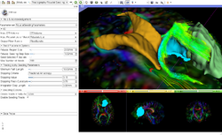
| + | *The [https://github.com/SlicerDMRI/slicerdmri.github.io/raw/master/docs/tutorials/FiberBundleSelectionAndScalarMeasurement.pdf Fiber Bundle Selection and Scalar Measurements] tutorial guides through the use of the Diffusion Bundle Selection module and the Fiber Tract Scalar Measurement module for diffusion MRI tractography data analysis. | ||
| + | *Author: Fan Zhang, University of Sydney Australia and Brigham and Women's Hospital | ||
| + | *Dataset: [[media:FiberBundleSelectionAndScalarMeasurement_TutorialContestWinter2016.zip| Fiber Bundle Selection And Scalar Measurement Tutorial Dataset]] | ||
| + | |align="right"| | ||
| + | [[File:FiberBundleSelectionAndScalarMeasurement_TutorialContestWinter2016_Snapshot.png|200px]] | ||
| + | |} | ||
| + | |||
| + | ===Plastimatch === | ||
| + | {|width="100%" | ||
| + | | | ||
| + | *The [http://www.na-mic.org/Wiki/images/5/5c/Plastimatch_TutorialContestWinter2016.pdf Plastimatch tutorial] guides through registration and wrapping of DICOM and DICOM-RT data using the Plastimatch extension of 3D Slicer. | ||
| + | *Author: Gregory Sharp, Massachusetts General Hospital | ||
| + | *Dataset: [http://www.na-mic.org/Wiki/index.php/File:Plastimatch_TutorialContestWinter2016.zip Plastimatch Tutorial Dataset] | ||
| + | |align="right"| | ||
| + | [[File:PlastimatchTutorial_Winter2016Contest.png|200px]] | ||
| + | |} | ||
| + | |||
| + | ===UKF === | ||
| + | {|width="100%" | ||
| + | | | ||
| + | *The [https://github.com/SlicerDMRI/slicerdmri.github.io/raw/master/docs/tutorials/UKFTractography.pdf UKF tutorial] guides through the use of the Unscented Kalman Filter (UKF) tractography module. | ||
| + | *Author: Pegah Kahali, Brigham and Women's Hopital | ||
| + | *Dataset: [http://www.na-mic.org/Wiki/index.php/File:UKF-Tractography_TutorialContestWinter2016.zip UKF tutorial Dataset] | ||
| + | |align="right"| | ||
| + | [[File:UKF_Winter2016.png|200px]] | ||
| + | |} | ||
| + | |||
| + | ==Summer 2014 Tutorial contest== | ||
| + | |||
| + | ===Cardiac Agatston Tutorial=== | ||
{|width="100%" | {|width="100%" | ||
| | | | ||
| Line 197: | Line 314: | ||
|} | |} | ||
| − | ==CMR Toolkit LA workflow== | + | ===CMR Toolkit LA workflow=== |
{|width="100%" | {|width="100%" | ||
| | | | ||
| Line 207: | Line 324: | ||
|} | |} | ||
| − | =Summer 2013 Tutorial contest= | + | ==Summer 2013 Tutorial contest== |
| − | ==Cardiac MRI Toolkit== | + | ===Cardiac MRI Toolkit=== |
{|width="100%" | {|width="100%" | ||
| | | | ||
| Line 219: | Line 336: | ||
|} | |} | ||
| − | ==HelloCLI== | + | ===HelloCLI=== |
{|width="100%" | {|width="100%" | ||
| | | | ||
| Line 229: | Line 346: | ||
|} | |} | ||
| − | ==SlicerRT== | + | ===SlicerRT=== |
{|width="100%" | {|width="100%" | ||
| | | | ||
| Line 239: | Line 356: | ||
|} | |} | ||
| − | ==DTIPrep== | + | ===DTIPrep=== |
{|width="100%" | {|width="100%" | ||
| | | | ||
| Line 249: | Line 366: | ||
|} | |} | ||
| − | = Summer 2012 Tutorial contest = | + | == Summer 2012 Tutorial contest == |
| − | ==Automatic Left Atrial Scar Segmenter == | + | ===Automatic Left Atrial Scar Segmenter === |
{|width="100%" | {|width="100%" | ||
| | | | ||
| Line 261: | Line 378: | ||
|} | |} | ||
| − | ==Qualitative and quantitative comparison of two RT dose distributions== | + | ===Qualitative and quantitative comparison of two RT dose distributions=== |
{|width="100%" | {|width="100%" | ||
| | | | ||
| Line 270: | Line 387: | ||
|} | |} | ||
| − | ==Dose accumulation for adaptive radiation therapy== | + | ===Dose accumulation for adaptive radiation therapy=== |
{|width="100%" | {|width="100%" | ||
| | | | ||
| Line 279: | Line 396: | ||
|} | |} | ||
| − | ==WebGL Export== | + | ===WebGL Export=== |
{|width="100%" | {|width="100%" | ||
| | | | ||
| Line 288: | Line 405: | ||
|} | |} | ||
| − | ==OpenIGTLink== | + | ===OpenIGTLink=== |
{|width="100%" | {|width="100%" | ||
| | | | ||
| Line 330: | Line 447: | ||
= External Resources = | = External Resources = | ||
| − | == Using the Editor == | + | == Murat Maga's blog posts about using 3D Slicer for biology == |
| + | |||
| + | * [https://blogs.uw.edu/maga/2017/04/11/getting-started-with-3d-slicer-as-a-biologist/ Slicer for Biologists] | ||
| + | * [https://blogs.uw.edu/maga/2017/04/11/a-worked-example-getting-and-visualizing-data-from-digimorph/ Loading data from DigiMorph] | ||
| + | * [https://blogs.uw.edu/maga/2017/04/11/morphosource-data-and-dealing-with-dicom-series-in-slicer/ Fixing problem DICOM] | ||
| + | * [https://blogs.uw.edu/maga/2017/04/12/scissors-tool-is-awesome/ Scissors tool is awesom] | ||
| + | |||
| + | == Using the (legacy) Editor == | ||
This set of tutorials about the use of slicer in paleontology is very well written and provides step-by-step instructions. Even though it covers slicer version 3.4, many of the concepts and techniques have applicability to the new version and to any 3D imaging field: | This set of tutorials about the use of slicer in paleontology is very well written and provides step-by-step instructions. Even though it covers slicer version 3.4, many of the concepts and techniques have applicability to the new version and to any 3D imaging field: | ||
Revision as of 12:08, 8 August 2017
Home < Documentation < Nightly < Training
|
For the latest Slicer documentation, visit the read-the-docs. |
Introduction: Slicer Nightly Tutorials
- This page contains "How to" tutorials with matched sample data sets. They demonstrate how to use the 3D Slicer environment (version Nightly release) to accomplish certain tasks.
- For tutorials for other versions of Slicer, please visit the Slicer training portal.
- For "reference manual" style documentation, please visit the Slicer Nightly documentation page
- For questions related to the Slicer4 Training Compendium, please send an e-mail to Sonia Pujol, Ph.D., Director of Training of 3D Slicer.
- Some of these tutorials are based on older releases of 3D Slicer. The concepts are still useful but bear in mind that some interface elements and features will be different in updated versions.
Contents
- 1 Introduction: Slicer Nightly Tutorials
- 2 General Introduction
- 3 Tutorials for software developers
- 4 Specific functions
- 4.1 Slicer4 Diffusion Tensor Imaging Tutorial
- 4.2 Slicer4 Neurosurgical Planning Tutorial
- 4.3 Slicer4 3D Visualization of DICOM images for Radiology Applications
- 4.4 Slicer4 Quantitative Imaging tutorial
- 4.5 Slicer4 IGT
- 4.6 Slicer4 3D Printing
- 4.7 Slicer4 Image Registration
- 4.8 Fast GrowCut
- 4.9 Slicer4 Radiation Therapy Tutorial
- 4.10 Other
- 5 3D Slicer Tutorial contests
- 6 Additional resources
- 7 External Resources
General Introduction
Slicer Welcome Tutorial
|
|

Slicer4Minute Tutorial
|
 |
Slicer4 Data Loading and 3D Visualization
|
 |
Tutorials for software developers
Slicer4 Programming Tutorial
|
 |
For additional Python scripts examples, please visit the Script Repository page
Developing and contributing extensions for 3D Slicer
|
 |
Specific functions
Slicer4 Diffusion Tensor Imaging Tutorial
|
 |
Slicer4 Neurosurgical Planning Tutorial
|
 |
Slicer4 3D Visualization of DICOM images for Radiology Applications
|
 |
Slicer4 Quantitative Imaging tutorial
|
 |
Slicer4 IGT
|
 |
Slicer4 3D Printing
|

|
Slicer4 Image Registration
|

|
- Based on: 3D Slicer version 4.5
See the Registration Library for worked out registration examples with data.
Fast GrowCut
|

|
Slicer4 Radiation Therapy Tutorial
- The SlicerRT tutorial is an introduction to the Radiation Therapy functionalities of Slicer.
- Author: Csaba Pinter, Andras Lasso, An Wang, Gregory C. Sharp, David Jaffray, Gabor Fichtinger.
- Dataset: download from MIDAS server
- Based on Slicer 4.7
Other
Additional (non-curated) videos-based demonstrations using 3D Slicer are accessible on You Tube.
3D Slicer Tutorial contests
Winter 2017 Tutorial contest
Segmentation for 3D printing
|
|

Slicer Pathology
|
|

Simple Python Tool for Quality Control of DWI data
|
|

SPHARM-PDM
|
|

Integration of Robot Operating System (ROS) and 3D Slicer using OpenIGTLink
|
|

Fiber Bundle Volume Measurement
|
|

Winter 2016 Tutorial contest
Subject Hierarchy
|
|

Fiber Bundle Selection and Scalar Measurements
|
|

Plastimatch
|
|

UKF
|
|

Summer 2014 Tutorial contest
Cardiac Agatston Tutorial
|
|

CMR Toolkit LA workflow
|
|

Summer 2013 Tutorial contest
Cardiac MRI Toolkit
|
|

HelloCLI
|
|
SlicerRT
|
|

DTIPrep
|
|

Summer 2012 Tutorial contest
Automatic Left Atrial Scar Segmenter
|
|

Qualitative and quantitative comparison of two RT dose distributions
|
|

Dose accumulation for adaptive radiation therapy
|
|

WebGL Export
|
|

OpenIGTLink
|
|

Additional resources
|

|
|

|
|
|
External Resources
Murat Maga's blog posts about using 3D Slicer for biology
Using the (legacy) Editor
This set of tutorials about the use of slicer in paleontology is very well written and provides step-by-step instructions. Even though it covers slicer version 3.4, many of the concepts and techniques have applicability to the new version and to any 3D imaging field:
- Open Source Paleontologist: 3D Slicer: The Tutorial
- Open Source Paleontologist: 3D Slicer: The Tutorial Part II
- Open Source Paleontologist: 3D Slicer: The Tutorial Part III
- Open Source Paleontologist: 3D Slicer: The Tutorial Part IV
- Open Source Paleontologist: 3D Slicer: The Tutorial Part V
- Open Source Paleontologist: 3D Slicer: The Tutorial Part VI
Team Contributions
See the collection of videos on the Kitware vimeo album.
User Contributions
See the User Contributions Page for more content.