Difference between revisions of "Documentation/Nightly/Modules/LASegmenter"
Liangjiazhu (talk | contribs) |
Liangjiazhu (talk | contribs) |
||
| Line 30: | Line 30: | ||
<!-- ---------------------------- --> | <!-- ---------------------------- --> | ||
{{documentation/{{documentation/version}}/module-section|Tutorials}} | {{documentation/{{documentation/version}}/module-section|Tutorials}} | ||
| − | + | *Load source image <br />[[file:LASegLoad.png]] | |
| − | [[file: | + | <br /> |
| + | |||
| + | *Selct the Editor module | ||
| + | ## Choose Master Volume and Merge Volume (label map to be used for the left atrium seed region) | ||
| + | ## Select PaintEffect and adjust Radius (in mm) so that the painted seed region (a circle) is completetly inside the left atrium | ||
| + | ## Go to the Axial view, choose a slice that is approximately to the center of the LA. Then, draw a circle that lies to the center of the left atrium on the slice | ||
| + | [[file:LASegSeedPanel.png|350px]][[file:LASegSeedImage.png]] | ||
| + | |||
| + | <br /> | ||
| + | *Go to the Segmentation->Left Atrium Segmentation module | ||
| + | [[file:LASegPanel.png|350px]][[file:LASegResult.png]] | ||
<!-- ---------------------------- --> | <!-- ---------------------------- --> | ||
Revision as of 02:53, 16 June 2013
Home < Documentation < Nightly < Modules < LASegmenter
|
For the latest Slicer documentation, visit the read-the-docs. |
Introduction and Acknowledgements
|
This work is part of the National Alliance for Medical Image Computing (NA-MIC), funded by the National Institutes of Health through the NIH Roadmap for Medical Research, Grant U54 EB005149. Information on NA-MIC can be obtained from the NA-MIC website. Contributor: LiangJia Zhu, University of Alabama at Birmingham | |||||||||
|


Tutorials
- Load source image
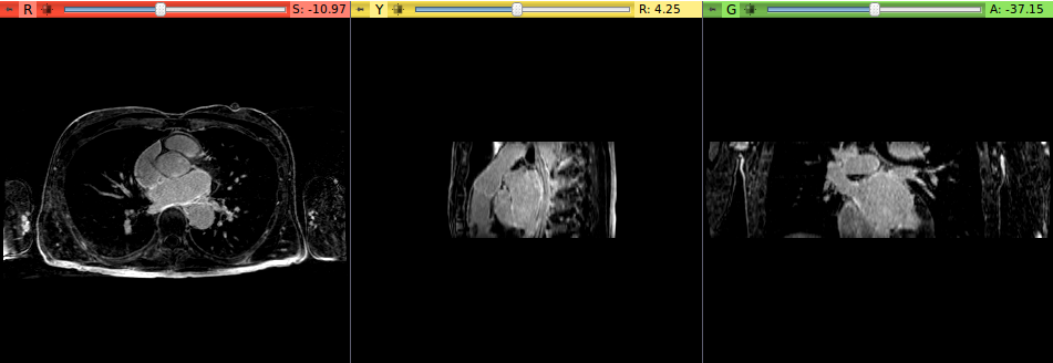

- Selct the Editor module
- Choose Master Volume and Merge Volume (label map to be used for the left atrium seed region)
- Select PaintEffect and adjust Radius (in mm) so that the painted seed region (a circle) is completetly inside the left atrium
- Go to the Axial view, choose a slice that is approximately to the center of the LA. Then, draw a circle that lies to the center of the left atrium on the slice


- Go to the Segmentation->Left Atrium Segmentation module


Panels and their use
NA
References
L. Zhu, Y. Gao, A. Yezzi, R. MacLeod, J. Cates, A. Tannenbaum. Automatic Segmentation of the Left Atrium from MRI Images Using Salient Feature and Contour Evolution, IEEE Engineering in Medicine and Biology Conference(EMBC), 2012.
Information for Developers
| Section under construction. |