Documentation/Nightly/Modules/AFTSegmenter
From Slicer Wiki
Revision as of 13:00, 25 July 2017 by Acsenrafilho (talk | contribs)
Home < Documentation < Nightly < Modules < AFTSegmenter

|
For the latest Slicer documentation, visit the read-the-docs. |
Introduction and Acknowledgements
|
Extension: LesionSpotlight | |||||||
|
Module Description
This module offers an implementation of a recent Multiple Sclerosis lesion segmentation approach based on a unsupervised method described by Cabezas et al. [1]. This module is intended to be used with FLAIR and T1 MRI volumes, which the MS lesions can be detected.
Use Cases
- Use Case 1: Multiple Sclerosis (MS) lesions segmentation
- Using T1 and FLAIR MRI volumes, it can be possible to detect abnormal voxel signal using a parametric strategy, which delineates white matter signals that does not belongs to the majority neighborhood pattern. More details can be found in the original paper [2]
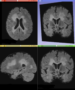
Input T2-FLAIR image with Multiple Sclerosis lesions
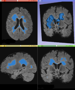
Lesion map resulted from AFT Segmenter module


Panels and their use

IO:
- T1 Volume
- Input T1 volume
- T2-FLAIR Volume
- Input T2-FLAIR volume
- Lesion Label
- Output a global lesion mask
- Is brain extracted?
- Is the input data (T1 and T2-FLAIR) already brain extracted?
Segmentation Parameters:
- Absolute Error Threshold
- Define the absolute error threshold for gray matter statistics. This measure evaluated the similarity between the MNI152 template and the T2-FLAIR gray matter fluctuation estimative. A higher error gives a higher variability in the final lesion segmentation
- Gamma
- Define the outlier detection based on units of standard deviation in the T2-FLAIR gray matter voxel intensity distribution
- White Matter Matching
- Set the local neighborhood searching for label refinement step. This metric defines the percentage of white matter tissue that surrounds the hyperintense lesions. Large values defines a conservative segmentation, i.e. in order to define a true MS lesion, it must be close to certain percentage of white matter area.
- Minimum Lesion Size
- Set the minimum lesion size adopted as a true lesion in the final lesion map. Units are given in number of voxels
- Gray Matter Mask Value
- Set the mask value that represents the gray matter. Default is defined based on the (Basic Brain Tissues module) output
- White Matter Mask Value
- Set the mask value that represents the white matter. Default is defined based on the (Basic Brain Tissues module) output
Similar Modules
References
- Cabezas, M., Oliver, A., Roura, E., Freixenet, J., Vilanova, J. C., Ramió-Torrentà, L., Rovira, À. and Lladó, X. (2014) ‘Automatic multiple sclerosis lesion detection in brain MRI by FLAIR thresholding’, Computer Methods and Programs in Biomedicine. Elsevier Ireland Ltd, 115(3), pp. 147–161. DOI: 10.1016/j.cmpb.2014.04.006.
Information for Developers
| Section under construction. |
- ↑ Cabezas M. et al.(2014) "Automatic multiple sclerosis lesion detection in brain MRI by FLAIR thresholding", Computer Methods and Programs in Biomedicine, DOI: 10.1016/j.cmpb.2014.04.006
- ↑ Cabezas M. et al.(2014) "Automatic multiple sclerosis lesion detection in brain MRI by FLAIR thresholding", Computer Methods and Programs in Biomedicine, DOI: 10.1016/j.cmpb.2014.04.006