Difference between revisions of "Documentation/Nightly/Extensions/LesionSpotlight"
Acsenrafilho (talk | contribs) m |
Acsenrafilho (talk | contribs) |
||
| Line 27: | Line 27: | ||
[[Image:LesionSpotlight-logo.png|left]] | [[Image:LesionSpotlight-logo.png|left]] | ||
| − | This extension provides image segmentation and enhancement approaches in order to increase the abnormal white matter voxels in magnetic resonance images. At moment, there are available the LS Segmenter | + | This extension provides a image segmentation and enhancement approaches in order to increase the abnormal white matter voxels in magnetic resonance images. At moment, there are available the '''LS Segmenter''' (specific for hyperintense Multiple Sclerosis lesion segmentation on T2-FLAIR images) and '''LS Contrast Enhancement''' (specific to increase the contrast of abnormal voxels of T2-FLAIR images) modules. The '''LS Contrast Enhancement''' module can be used as an image enhancement pre-processing step before a future lesion segmentation algorithm (which could be used any other approach, such as LesionTOADS, OASIS or LST). The '''LS Segmenter''' module implements a hyperintense T2-FLAIR lesion segmentation based on the algorithm published in the paper<ref>DOI:</ref>. |
| − | '''NOTE''': The Logistic Contrast Enhancement, Weighted Enhancement Image Filter and Automatic FLAIR Threshold modules are only supporting CLI methods added | + | '''NOTE''': The Logistic Contrast Enhancement, Weighted Enhancement Image Filter and Automatic FLAIR Threshold modules are only supporting CLI methods added in the extension to calculate specific parts of the segmentation procedure. For this reason, these modules are '''not''' supposed to be used alone. Please, use the '''LS Contrast Enhancer''' or '''LS Segmenter''' modules only. |
<!-- ---------------------------- --> | <!-- ---------------------------- --> | ||
| Line 40: | Line 40: | ||
Most frequently used for these scenarios: | Most frequently used for these scenarios: | ||
* Use Case 1: Multiple Sclerosis (MS) hyperintense lesions segmentation | * Use Case 1: Multiple Sclerosis (MS) hyperintense lesions segmentation | ||
| − | **Hyperintense Multiple Sclerosis lesions, mainly in T2-FLAIR images, have | + | **Hyperintense Multiple Sclerosis lesions, mainly in T2-FLAIR images, have an easy application of this segmentations procedure. |
* Use Case 2: Increase contrast in abnormal voxels in white matter | * Use Case 2: Increase contrast in abnormal voxels in white matter | ||
| − | **There are some lesion segmentation approaches that relies on the voxel intensity level presented in the lesion signal, where the LS Contrast Enhancer module can be helpful to increase the contrast between lesions and surrounding brain tissues (mainly | + | **There are some lesion segmentation approaches that relies on the voxel intensity level presented in the lesion signal, where the '''LS Contrast Enhancer''' module can be helpful to increase the contrast between lesions and surrounding brain tissues (mainly normal appearing white matter - NAWM). |
<gallery widths="300px" heights="300px" perrow="3"> | <gallery widths="300px" heights="300px" perrow="3"> | ||
Revision as of 18:46, 16 February 2017
Home < Documentation < Nightly < Extensions < LesionSpotlight
|
For the latest Slicer documentation, visit the read-the-docs. |
Introduction and Acknowledgements
|
This work was partially funded by CAPES and CNPq, a Brazillian Agencies. Information on CAPES can be obtained on the CAPES website and CNPq website. | |||||||||
|
Extension Description
This extension provides a image segmentation and enhancement approaches in order to increase the abnormal white matter voxels in magnetic resonance images. At moment, there are available the LS Segmenter (specific for hyperintense Multiple Sclerosis lesion segmentation on T2-FLAIR images) and LS Contrast Enhancement (specific to increase the contrast of abnormal voxels of T2-FLAIR images) modules. The LS Contrast Enhancement module can be used as an image enhancement pre-processing step before a future lesion segmentation algorithm (which could be used any other approach, such as LesionTOADS, OASIS or LST). The LS Segmenter module implements a hyperintense T2-FLAIR lesion segmentation based on the algorithm published in the paper[1].
NOTE: The Logistic Contrast Enhancement, Weighted Enhancement Image Filter and Automatic FLAIR Threshold modules are only supporting CLI methods added in the extension to calculate specific parts of the segmentation procedure. For this reason, these modules are not supposed to be used alone. Please, use the LS Contrast Enhancer or LS Segmenter modules only.
Modules
Use Cases
Most frequently used for these scenarios:
- Use Case 1: Multiple Sclerosis (MS) hyperintense lesions segmentation
- Hyperintense Multiple Sclerosis lesions, mainly in T2-FLAIR images, have an easy application of this segmentations procedure.
- Use Case 2: Increase contrast in abnormal voxels in white matter
- There are some lesion segmentation approaches that relies on the voxel intensity level presented in the lesion signal, where the LS Contrast Enhancer module can be helpful to increase the contrast between lesions and surrounding brain tissues (mainly normal appearing white matter - NAWM).

T2-FLAIR image from a MS patient
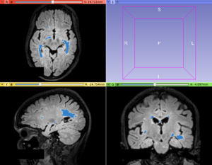
T2-FLAIR hyperintense lesion segmentation provided by the LS Segmenter module
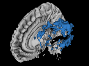
Lesion and White Matter 3D representation
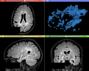
T2-FLAIR image with original lesion contrast
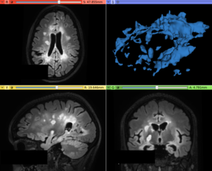
T2-FLAIR image with hyperintense lesion contrast enhanced provided by the LS Contrast Enhancer module


Similar Extensions
N/A
References
- paper
Information for Developers
| Section under construction. |
Repositories:
- Source code: GitHub repository
- Issue tracker: open issues and enhancement requests
- ↑ DOI: