Difference between revisions of "Documentation/Nightly/Extensions/DiffusionComplexityMap"
Acsenrafilho (talk | contribs) m (Infos addition) |
Acsenrafilho (talk | contribs) m Tag: 2017 source edit |
||
| Line 22: | Line 22: | ||
<!-- ---------------------------- --> | <!-- ---------------------------- --> | ||
| − | {{documentation/{{documentation/version}}/module-section| | + | {{documentation/{{documentation/version}}/module-section|Module Description}} |
| − | + | This module offer a simple application to the Anisotropic Anomalous Diffusion (AAD) filter, which is able to increase the image SNR and preserve fine object's details around the image space. This method was studied on MRI structural images (T1 and T2), which other imaging modalities could be properly investigated in the future. | |
| − | |||
| − | |||
| − | |||
| − | |||
| − | |||
| − | |||
| − | |||
| − | |||
| − | |||
| − | |||
| − | |||
| − | |||
| − | |||
<!-- ---------------------------- --> | <!-- ---------------------------- --> | ||
{{documentation/{{documentation/version}}/module-section|Use Cases}} | {{documentation/{{documentation/version}}/module-section|Use Cases}} | ||
| − | + | * Use Case 1: Noise reduction as a preprocessing step for tissue segmentation | |
| − | * Use Case 1: Noise reduction as a | + | **When dealing with single voxel classification schemes running noise reduction as a preprocessing scheme will reduce the number of single misclassified voxels. |
| − | **When dealing with single voxel classification schemes | + | * Use Case 2: Preprocessing to volume rendering |
| − | * Use Case 2: | ||
**Noise reduction will result in nicer looking volume renderings | **Noise reduction will result in nicer looking volume renderings | ||
* Use Case 3: Noise reduction as part of image processing pipeline | * Use Case 3: Noise reduction as part of image processing pipeline | ||
**Could offer a better segmentation and classification on specific brain image analysis such as in Multiple Sclerosis lesion segmentation | **Could offer a better segmentation and classification on specific brain image analysis such as in Multiple Sclerosis lesion segmentation | ||
| − | <gallery widths=" | + | |
| + | <gallery widths="300px" perrow="3"> | ||
Image:MRI_raw.png|Raw T1 weighted MRI Image | Image:MRI_raw.png|Raw T1 weighted MRI Image | ||
Image:MRI_AAD.png|T1 weighted MRI Image with AAD filter (q=1.2) | Image:MRI_AAD.png|T1 weighted MRI Image with AAD filter (q=1.2) | ||
| − | |||
| − | |||
| − | |||
</gallery> | </gallery> | ||
<!-- ---------------------------- --> | <!-- ---------------------------- --> | ||
| − | {{documentation/{{documentation/version}}/ | + | {{documentation/{{documentation/version}}/module-section|Panels and their use}} |
| − | *[[Documentation/{{documentation/version}}/Modules/ | + | |
| + | [[Image:aad_scalar_gui.png|thumb|380px|User Interface]] | ||
| + | '''IO:''' | ||
| + | *'''Input Volume''' | ||
| + | **Select the input image | ||
| + | *'''Output Volume''' | ||
| + | **Set the output image file which the filters should place the final result | ||
| + | |||
| + | '''Diffusion Parameters:''' | ||
| + | *'''Conductance''' | ||
| + | **The conductance regulates the diffusion intensity in the neighborhood area. Choose a higher conductance if the input image has strong noise seem in the whole image space. | ||
| + | *'''Use Auto Conductance''' | ||
| + | **Choose if you want to use an automatic adjustment of conductance parameter. If this is checked, the inserted value is ignored and the optimization function below is used. | ||
| + | *'''Optimization Function''' | ||
| + | **A set of optimization function for automatic estimation of conductance parameter. This is helpful is you do not have an initial guess on what value is appropriate to the conductance setting. (Canny, MAD and Morphological). Please see the [http://www.insight-journal.org/browse/publication/983 Insight-Journal article] that explain each of these automatic conductance adjustment methods. | ||
| + | *'''Number of Iteractions''' | ||
| + | **The number of iterations regulates the numerical simulation of the anomalous process over the image. This parameters is also related with the de-noising intensity, however it is more sensible to the noise intensity. Choose the higher number of iterations if the image presents high intensity noise which is not well treated by the conductance parameter. | ||
| + | *'''Time Step''' | ||
| + | **The time step is a normalization parameters for the numerical simulation. The maximum value, given as default, is set to 3D images. Lower time step restrict the numerical simulation of the anomalous process. | ||
| + | *'''Anomalous parameter''' | ||
| + | **The anomalous parameter (or q value) is the generalization parameters responsible to give the anomalous process approach on the diffusion equation. See the reference paper<ref>Da S Senra Filho, A. C., Garrido Salmon, C. E., & Murta Junior, L. O. (2015). Anomalous diffusion process applied to magnetic resonance image enhancement. Physics in Medicine and Biology, 60(6), 2355–2373. doi:10.1088/0031-9155/60/6/2355</ref> to choose the appropriate q value (at moment, only tested in MRI T1 and T2 weighted images). | ||
| + | |||
| + | |||
| + | <!-- ---------------------------- --> | ||
| + | {{documentation/{{documentation/version}}/module-section|Similar Modules}} | ||
| + | *[[Documentation/{{documentation/version}}/Modules/IADImageFilter|IAD Image Filter]] TODO Colocar outros links | ||
<!-- ---------------------------- --> | <!-- ---------------------------- --> | ||
{{documentation/{{documentation/version}}/extension-section|References}} | {{documentation/{{documentation/version}}/extension-section|References}} | ||
| − | * | + | * |
| − | |||
| − | |||
| − | |||
| − | |||
<!-- ---------------------------- --> | <!-- ---------------------------- --> | ||
| Line 73: | Line 78: | ||
Repositories: | Repositories: | ||
| − | *Source code: [https://github.com/CSIM-Toolkits/ | + | *Source code: [https://github.com/CSIM-Toolkits/SlicerDiffusionComplexityMap GitHub repository] |
| − | *Issue tracker: [https://github.com/CSIM-Toolkits/ | + | *Issue tracker: [https://github.com/CSIM-Toolkits/SlicerDiffusionComplexityMap/issues open issues and enhancement requests] |
<!-- ---------------------------- --> | <!-- ---------------------------- --> | ||
Revision as of 12:36, 12 March 2024
Home < Documentation < Nightly < Extensions < DiffusionComplexityMap
|
For the latest Slicer documentation, visit the read-the-docs. |
Introduction and Acknowledgements
|
This work was funded by University of Campinas, Brazil. More information on the website Unicamp website. | |||||||||
|
Module Description
This module offer a simple application to the Anisotropic Anomalous Diffusion (AAD) filter, which is able to increase the image SNR and preserve fine object's details around the image space. This method was studied on MRI structural images (T1 and T2), which other imaging modalities could be properly investigated in the future.
Use Cases
- Use Case 1: Noise reduction as a preprocessing step for tissue segmentation
- When dealing with single voxel classification schemes running noise reduction as a preprocessing scheme will reduce the number of single misclassified voxels.
- Use Case 2: Preprocessing to volume rendering
- Noise reduction will result in nicer looking volume renderings
- Use Case 3: Noise reduction as part of image processing pipeline
- Could offer a better segmentation and classification on specific brain image analysis such as in Multiple Sclerosis lesion segmentation

Raw T1 weighted MRI Image
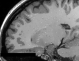
T1 weighted MRI Image with AAD filter (q=1.2)


Panels and their use

IO:
- Input Volume
- Select the input image
- Output Volume
- Set the output image file which the filters should place the final result
Diffusion Parameters:
- Conductance
- The conductance regulates the diffusion intensity in the neighborhood area. Choose a higher conductance if the input image has strong noise seem in the whole image space.
- Use Auto Conductance
- Choose if you want to use an automatic adjustment of conductance parameter. If this is checked, the inserted value is ignored and the optimization function below is used.
- Optimization Function
- A set of optimization function for automatic estimation of conductance parameter. This is helpful is you do not have an initial guess on what value is appropriate to the conductance setting. (Canny, MAD and Morphological). Please see the Insight-Journal article that explain each of these automatic conductance adjustment methods.
- Number of Iteractions
- The number of iterations regulates the numerical simulation of the anomalous process over the image. This parameters is also related with the de-noising intensity, however it is more sensible to the noise intensity. Choose the higher number of iterations if the image presents high intensity noise which is not well treated by the conductance parameter.
- Time Step
- The time step is a normalization parameters for the numerical simulation. The maximum value, given as default, is set to 3D images. Lower time step restrict the numerical simulation of the anomalous process.
- Anomalous parameter
- The anomalous parameter (or q value) is the generalization parameters responsible to give the anomalous process approach on the diffusion equation. See the reference paper[1] to choose the appropriate q value (at moment, only tested in MRI T1 and T2 weighted images).
Similar Modules
- IAD Image Filter TODO Colocar outros links
References
Information for Developers
| Section under construction. |
Repositories:
- Source code: GitHub repository
- Issue tracker: open issues and enhancement requests
- ↑ Da S Senra Filho, A. C., Garrido Salmon, C. E., & Murta Junior, L. O. (2015). Anomalous diffusion process applied to magnetic resonance image enhancement. Physics in Medicine and Biology, 60(6), 2355–2373. doi:10.1088/0031-9155/60/6/2355