Home < Documentation < Nightly < Modules < SUVFactorCalculator
Introduction and Acknowledgements
|
Acknowledgments:
This work is funded in part by Quantitative Imaging to Assess Response in Cancer Therapy Trials NIH grant U01-CA140206 and Quantitative Image Informatics for Cancer Research (QIICR) NIH grant U24 CA180918.
Authors: Andrey Fedorov (SPL), Ethan Ulrich (University of Iowa), Markus van Tol (University of Iowa)
Contributors: Christian Bauer (University of Iowa), Reinhard Beichel (University of Iowa), John Buatti (University of Iowa), Hans Johnson (University of Iowa), Wendy Plesniak (SPL, BWH), Nicole Aucoin (SPL, BWH), Ron Kikinis (SPL, BWH)
Contact: <email>qin@iibi.uiowa.edu</email>
License: Slicer License
|
| The University of Iowa (UIowa)
|
| Quantitative Image Informatics for Cancer Research
|
| National Alliance for Medical Image Computing (NA-MIC)
|
|
|
General Information
Module Type & Category
Type: CLI
Category: Quantification
Module Description
| Program title |
SUV Factor Calculator
|
| Program description |
This command line interface module takes a PET DICOM series and creates a Real World Value Mapping (RWVM) file including 4 types of Standardized Uptake Value conversion factors (SUVbw, SUVlbm, SUVbsa, SUVibw).
|
| Program documentation-url |
Source code on GitHub
|
Usage
Use Cases, Examples
The patient size correction factors are summarized here, where weight is in kilograms and height is in centimeters.
- SUVbw:
- SUVlbm:
- males: 1.10 * weight - 128 * (weight/height)^2
- females: 1.07 * weight - 148 * (weight/height)^2
- SUVbsa:
- males & females: weight^0.425 * height^0.725 * 0.007184
- SUVibw:
- males: 48.0 + 1.06 * (height - 152)
- females: 45.5 + 0.91 * (height - 152)
Tutorials
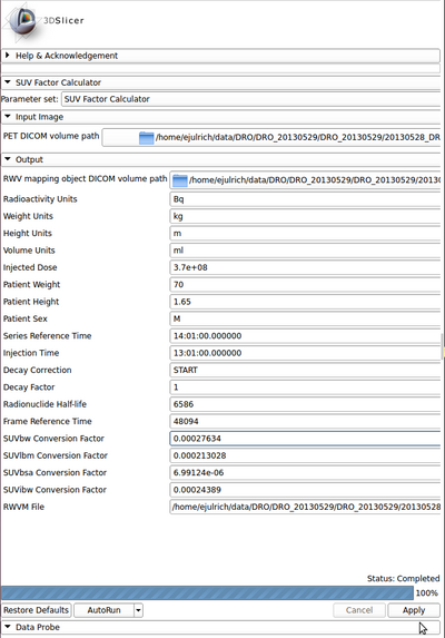
Quick Tour of Features and Use
A list of panels in the interface, their features, what they mean, and how to use them.
- Input Image:
- PET DICOM volume path [----petDICOMPath] [--p]: Input path to a directory containing a PET DICOM series with header information for SUV computation
- Output:
- RWV mapping object DICOM volume path [----rwvmDICOMPath] [--r]: Output path to a directory to store the RWV object with the SUV computation result
- Radioactivity Units : DICOM Tag (0054,1001) - Radioactivity Units
- Weight Units : Patient weight units (always assumed to be kg)
- Height Units : Patient weight units (always assumed to be meters)
- Volume Units : Volume units for concentration (always assumed to be mL)
- Injected Dose : DICOM Tag (0018,1074) - Radionuclide Total Dose
- Patient Weight : DICOM Tag (0010,1030) - Patient Weight
- Patient Height : DICOM Tag (0010,1020) - Patient Size
- Patient Sex : DICOM Tag (0010,0040) - Patient Sex
- Series Reference Time : DICOM Tag (0008,0031) - Series Time
- Injection Time : DICOM Tag (0018,1072) - Radiopharmaceutical Start Time
- Decay Correction : DICOM Tag (0054,1102) - Decay Correction
- Decay Factor : DICOM Tag (0054,1321) - Decay Factor
- Radionuclide Half-life : DICOM Tag (0018,1075) - Radionuclide Half-Life
- Frame Reference Time : DICOM Tag (0054,1300) - Frame Reference Time
- SUVbw Conversion Factor : Standardized Uptake Value (body weight) Conversion Factor
- SUVlbm Conversion Factor : Standardized Uptake Value (lean body mass) Conversion Factor
- SUVbsa Conversion Factor : Standardized Uptake Value (body surface area) Conversion Factor
- SUVibw Conversion Factor : Standardized Uptake Value (ideal body weight) Conversion Factor
- RWVM File : Real World Value Mapping object file
|
|
Development
Information for Developers
References
Sugawara et al. "Reevaluation of the Standardized Uptake Value for FDG: Variation with Body Weight and Methods for Correction." Radiology. 1999.
http://pubs.rsna.org/doi/pdf/10.1148/radiology.213.2.r99nv37521


