Main Page/SlicerCommunity
Contents
- 1 3D Slicer Enabled Research
- 2 2017
- 2.1 A Study of Volumetric Variations of Basal Nuclei in the Normal Human Brain by Magnetic Resonance Imaging
- 2.2 Associations of Radiomic Data Extracted from Static and Respiratory-Gated CT Scans with Disease Recurrence in Lung Cancer Patients Treated with SBRT
- 2.3 Hybrid Positron Emission Tomography Segmentation of Heterogeneous Lung Tumors using 3D Slicer: Improved Growcut Algorithm with Threshold Initialization
- 2.4 Pre-clinical Validation of Virtual Bronchoscopy using 3D Slicer
- 2.5 Theoretical Observation on Diagnosis Maneuver for Benign Paroxysmal Positional Vertigo
- 2.6 Anatomical Study and Locating Nasolacrimal Duct on Computed Topographic Image
- 2.7 Intra-rater Variability in Low-grade Glioma Segmentation
- 2.8 Biomaterial Shell Bending with 3D-printed Templates in Vertical and Alveolar Ridge Augmentation: A technical Note
3D Slicer Enabled Research
3D Slicer is a free open source software package distributed under a BSD style license. The majority of funding for the development of 3D slicer comes from a number of grants and contracts from the National Institutes of Health. See Slicer Acknowledgments for more information.
This page focuses on research that was done outside of our immediate collaboration community. That community is represented in the publication database.
We invite you to provide information on how you are using 3D Slicer to produce peer-reviewed research. Information about the scientific impact of this tool is helpful in raising funding for the continued support.
2016:: 2015 :: 2014-2011 :: 2010-2005
2017
A Study of Volumetric Variations of Basal Nuclei in the Normal Human Brain by Magnetic Resonance Imaging
|
Publication: Clin Anat. 2017 Mar;30(2):175-82. PMID: 28078760 Authors: Elkattan A, Mahdy A, Eltomey M, Ismail R. Institution: Department of Anatomy, Tanta University of Medical Sciences, Tanta, Egypt. Background/Purpose: Knowledge of the effects of healthy aging on brain structures is necessary to identify abnormal changes due to diseases. Many studies have demonstrated age-related volume changes in the brain using MRI. 60 healthy individuals who had normal MRI aged from 20 years to 80 years were examined and classified into three groups: Group I: 21 persons; nine males and 12 females aging between 20-39 years old. Group II: 22 persons; 11 males and 11 females aging between 40-59 years old. Group III: 17 persons; eight males and nine females aging between 60-80 years old. Volumetric analysis was done to evaluate the effect of age, gender and hemispheric difference in the caudate and putamen by the 3D Slicer 4.3.3.1 software using 3D T1-weighted images. Data were analyzed by student's unpaired t test, ANOVA and regression analysis. The volumes of the measured and corrected caudate nuclei and putamen significantly decreased with aging in males. There was a statistically insignificant relation between the age and the volume of the measured caudate nuclei and putamen in females but there was a statistically significant relation between the age and the corrected caudate nuclei and putamen. There was no significant difference on the caudate and putamen volumes between males and females. There was no significant difference between the right and left caudate nuclei volumes. There was a leftward asymmetry in the putamen volumes. The results can be considered as a base to track individual changes with time (aging and CNS diseases). |
Associations of Radiomic Data Extracted from Static and Respiratory-Gated CT Scans with Disease Recurrence in Lung Cancer Patients Treated with SBRT
|
Publication: PLoS One. 2017 Jan 3;12(1):e0169172. PMID: 28046060| PDF Authors: Huynh E, Coroller TP, Narayan V, Agrawal V, Romano J, Franco I, Parmar C, Hou Y, Mak RH, Aerts HJ. Institution: Department of Radiation Oncology, Dana-Farber Cancer Institute, Brigham and Women's Hospital, Harvard Medical School, Boston, USA. Background/Purpose: Radiomics aims to quantitatively capture the complex tumor phenotype contained in medical images to associate them with clinical outcomes. This study investigates the impact of different types of computed tomography (CT) images on the prognostic performance of radiomic features for disease recurrence in early stage non-small cell lung cancer (NSCLC) patients treated with stereotactic body radiation therapy (SBRT). 112 early stage NSCLC patients treated with SBRT that had static free breathing (FB) and average intensity projection (AIP) images were analyzed. Nineteen radiomic features were selected from each image type (FB or AIP) for analysis based on stability and variance. The selected FB and AIP radiomic feature sets had 6 common radiomic features between both image types and 13 unique features. The prognostic performances of the features for distant metastasis (DM) and locoregional recurrence (LRR) were evaluated using the concordance index (CI) and compared with two conventional features (tumor volume and maximum diameter). P-values were corrected for multiple testing using the false discovery rate procedure. None of the FB radiomic features were associated with DM, however, seven AIP radiomic features, that described tumor shape and heterogeneity, were (CI range: 0.638-0.676). Conventional features from FB images were not associated with DM, however, AIP conventional features were (CI range: 0.643-0.658). Radiomic and conventional multivariate models were compared between FB and AIP images using cross validation. The differences between the models were assessed using a permutation test. AIP radiomic multivariate models (median CI = 0.667) outperformed all other models (median CI range: 0.601-0.630) in predicting DM. None of the imaging features were prognostic of LRR. Therefore, image type impacts the performance of radiomic models in their association with disease recurrence. AIP images contained more information than FB images that were associated with disease recurrence in early stage NSCLC patients treated with SBRT, which suggests that AIP images may potentially be more optimal for the development of an imaging biomarker. Funding:
|
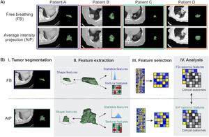
 A) Examples of free breathing (FB) and average intensity projection (AIP) images, demonstrating the observable differences in tumor phenotype between each image type. AIP images were reconstructed from 4D computed tomography (CT) scans. B) Schematic representation of the radiomics workflow for FB and AIP images. I. CT images of the patient are acquired and the tumor is segmented. II. Imaging features (radiomic and conventional features) are extracted from the tumor volume. III. Radiomic features undergo a feature dimension reduction process to generate a low-dimensional feature set based on feature stability and variance. IV. Imaging features are then analyzed with clinical outcomes to evaluate their prognostic power. FB and AIP radiomics features are compared. A set of 644 radiomic features was extracted from tumor volumes isolated from FB or AIP images (Fig 1B) using an in-house Matlab 2013 toolbox and 3D Slicer 4.4.0 software] |
Hybrid Positron Emission Tomography Segmentation of Heterogeneous Lung Tumors using 3D Slicer: Improved Growcut Algorithm with Threshold Initialization
|
Publication: J. Med. Imag. 2017 Jan-Mar;4(1), 011009. PMID: 28149920 | PDF Authors: Hannah Mary T. Thomas, Devadhas Devakumar, Balukrishna Sasidharan, Stephen R. Bowen, Danie Kingslin Heck, E. James Jebaseelan Samuel Institution: VIT University, School of Advanced Sciences, Department of Physics, Vellore, Tamil Nadu 632004, India. Background/Purpose: This paper presents an improved GrowCut (IGC), a positron emission tomography-based segmen- tation algorithm, and tests its clinical applicability. Contrary to the traditional method that requires the user to provide the initial seeds, the IGC algorithm starts with a threshold-based estimate of the tumor and a three- dimensional morphologically grown shell around the tumor as the foreground and background seeds, respec- tively. The repeatability of IGC from the same observer at multiple time points was compared with the traditional GrowCut algorithm. The algorithm was tested in 11 nonsmall cell lung cancer lesions and validated against the clinician-defined manual contour and compared against the clinically used 25% of the maximum standardized uptake value [SUV-(max)], 40% SUVmax, and adaptive threshold methods. The time to edit IGC-defined functional volume to arrive at the gross tumor volume (GTV) was compared with that of manual contouring. The repeatability of the IGC algorithm was very high compared with the traditional GrowCut (p = 0.003) and demonstrated higher agreement with the manual contour with respect to threshold-based methods. Compared with manual contouring, editing the IGC achieved the GTV in significantly less time (p = 0.11). The IGC algorithm offers a highly repeatable functional volume and serves as an effective initial guess that can well minimize the time spent on labor-intensive manual contouring. |
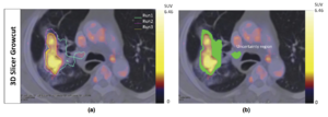
 A) A representative example of the uncertainty volume observed with the 3D Slicer GrowCutmethod. (a) The lesion was delineated in three separate runs. There was variability with each run and the composite error in the variability calculated as the uncertainty volume is highlighted in green in (b). |
Pre-clinical Validation of Virtual Bronchoscopy using 3D Slicer
|
Publication: Int J Comput Assist Radiol Surg. 2017 Jan;12(1):25-38. PMID: 27325238 Authors: Nardelli P, Jaeger A, O'Shea C, Khan KA, Kennedy MP, Cantillon-Murphy P. Institution: School of Engineering, University College Cork, College Road, Cork, Ireland. Background/Purpose: Lung cancer still represents the leading cause of cancer-related death, and the long-term survival rate remains low. Computed tomography (CT) is currently the most common imaging modality for lung diseases recognition. The purpose of this work was to develop a simple and easily accessible virtual bronchoscopy system to be coupled with a customized electromagnetic (EM) tracking system for navigation in the lung and which requires as little user interaction as possible, while maintaining high usability. Methods: The proposed method has been implemented as an extension to the open-source platform, 3D Slicer. It creates a virtual reconstruction of the airways starting from CT images for virtual navigation. It provides tools for pre-procedural planning and virtual navigation, and it has been optimized for use in combination with a [Formula: see text] of freedom EM tracking sensor. Performance of the algorithm has been evaluated in ex vivo and in vivo testing. Results: During ex vivo testing, nine volunteer physicians tested the implemented algorithm to navigate three separate targets placed inside a breathing pig lung model. In general, the system proved easy to use and accurate in replicating the clinical setting and seemed to help choose the correct path without any previous experience or image analysis. Two separate animal studies confirmed technical feasibility and usability of the system. Conclusions: This work describes an easily accessible virtual bronchoscopy system for navigation in the lung. The system provides the user with a complete set of tools that facilitate navigation towards user-selected regions of interest. Results from ex vivo and in vivo studies showed that the system opens the way for potential future work with virtual navigation for safe and reliable airway disease diagnosis. |
Theoretical Observation on Diagnosis Maneuver for Benign Paroxysmal Positional Vertigo
|
Publication: Acta Otolaryngol. 2017 Jan 13:1-8. PMID: 28084876 Authors: Yang XK, Zheng YY, Yang XG. Institution: Neurology Department , Wenzhou People's Hospital , Wenzhou , Zhejiang , PR China. Background/Purpose: To make a comprehensive analysis with a variety of diagnostic maneuvers is conducive to the correct diagnosis and classification of BPPV. OBJECTIVE: Based on the standard spatial coordinate-based semicircular canal model for theoretical observation on diagnostic maneuvers for benign paroxysmal positional vertigo (BPPV) to analyze the meaning and key point of each step of the maneuver. MATERIALS AND METHODS: This study started by building a standard model of semicircular canal with space orientation by segmentation of the inner ear done with the 3D Slicer software based on MRI scans, then gives a demonstration and observation of BPPV diagnostic maneuvers by using the model. RESULTS: The supine roll maneuver is mainly for diagnosis of lateral semicircular canal BPPV. The Modified Dix-Hallpike maneuver is more specific for the diagnosis of posterior semicircular canal BPPV. The side-lying bow maneuver designed here is theoretically suitable for diagnosis of anterior semicircular canal BPPV. |
Anatomical Study and Locating Nasolacrimal Duct on Computed Topographic Image
|
Publication: J Craniofac Surg. 2017 Jan;28(1):275-79. PMID: 27977487 Authors: Zhang S, Cheng Y, Xie J, Wang Z, Zhang F, Chen L, Feng Y, Wang G. Institution: Department of Endocrine †Department of Neurosurgery, First Hospital of Jilin University, Changchun, China. Background/Purpose: We performed a novel anatomical and radiological investigation to understand the structure of nasolacrimal duct (NLD) and to provide data to help surgeons locate the openings of NLD efficiently based on landmarks. MATERIALS AND METHODS: We examined the NLD region using computed tomography images of 133 individuals and 6 dry skull specimens. Multiplanar reconstruction of the computed tomography images was performed, and the anatomical features of the NLD were studied in the coronal, sagittal, and axial planes. The long and short diameters of NLD were measured along its cross-section. The position of NLD was localized using the nostril, concha nasalis media, and medial orbital corner as landmarks. The free and open source software, 3D Slicer, was used for the segmentation of the NLD and 3D visualization of the superior and inferior openings of the NLD. RESULTS: The length, angle, and diameter of NLD were significantly influenced by the age in females compared to those in males. The inferior opening of the NLD could be located efficiently using the nostril and the midsagittal line while the superior opening of NLD could be located using the medial orbital corner. Third, 3D Slicer enabled us to measure the distance between the skin and the bony structure in the image. CONCLUSION: Our study indicates that the sex and age of the patient should be considered while selecting the optimal NLD stent for a patient, and that the precise location of NLD in reference to landmarks can simplify the surgical difficulties and reduce the risk of injury during the transnasal operation. |
Intra-rater Variability in Low-grade Glioma Segmentation
|
Publication: J Neurooncol. 2017 Jan;131(2):393-402. PMID: 27837437 Authors: Bø HK, Solheim O, Jakola AS, Kvistad KA, Reinertsen I, Berntsen EM. Institution: Department of Radiology and Nuclear Medicine, St. Olavs University Hospital, Trondheim, Norway Background/Purpose: Assessment of size and growth are key radiological factors in low-grade gliomas (LGGs), both for prognostication and treatment evaluation, but the reliability of LGG-segmentation is scarcely studied. With a diffuse and invasive growth pattern, usually without contrast enhancement, these tumors can be difficult to delineate. The aim of this study was to investigate the intra-observer variability in LGG-segmentation for a radiologist without prior segmentation experience. Pre-operative 3D FLAIR images of 23 LGGs were segmented three times in the software 3D Slicer. Tumor volumes were calculated, together with the absolute and relative difference between the segmentations. To quantify the intra-rater variability, we used the Jaccard coefficient comparing both two (J2) and three (J3) segmentations as well as the Hausdorff Distance (HD). The variability measured with J2 improved significantly between the two last segmentations compared to the two first, going from 0.87 to 0.90 (p = 0.04). Between the last two segmentations, larger tumors showed a tendency towards smaller relative volume difference (p = 0.07), while tumors with well-defined borders had significantly less variability measured with both J2 (p = 0.04) and HD (p < 0.01). We found no significant relationship between variability and histological sub-types or Apparent Diffusion Coefficients (ADC). We found that the intra-rater variability can be considerable in serial LGG-segmentation, but the variability seems to decrease with experience and higher grade of border conspicuity. Our findings highlight that some criteria defining tumor borders and progression in 3D volumetric segmentation is needed, if moving from 2D to 3D assessment of size and growth of LGGs. |
Biomaterial Shell Bending with 3D-printed Templates in Vertical and Alveolar Ridge Augmentation: A technical Note
|
Publication: Oral Surg Oral Med Oral Pathol Oral Radiol. 2017 Jan 4. PMID: 28215503 Authors: Draenert FG, Gebhart F, Mitov G, Neff A. Institution: Oral & Maxillofacial Surgery, University of Marburg, Germany. Background/Purpose:
Alveolar ridge and vertical augmentations are challenging procedures in dental implantology. Even material blocks with an interconnecting porous system are never completely resorbed. Shell techniques combined with autologous bone chips are therefore the gold standard. Using biopolymers for these techniques is well documented. We applied three-dimensional (3-D) techniques to create an individualized bending model for the adjustment of a plane biopolymer membrane made of polylactide.
|