What is 3D Slicer ?
Desktop software to solve advanced image computing challenges with a focus on clinical and biomedical applications.
Development platform to quickly build and deploy custom solutions for research and commercial products, using free, open source software.
Community of knowledgeable users and developers working together to improve medical computing.
Development platform to quickly build and deploy custom solutions for research and commercial products, using free, open source software.
Community of knowledgeable users and developers working together to improve medical computing.
See 3D Slicer in action
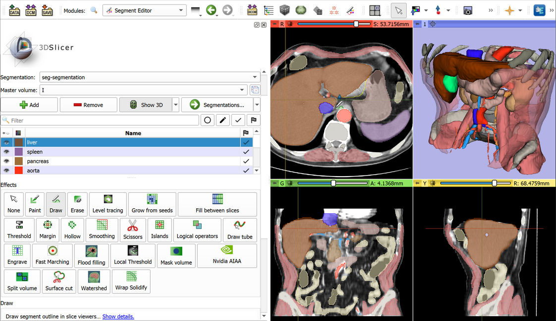
Image segmentation
Create surgical plans, create high-quality atlases, or training data sets for deep learning using the Segment Editor module. learn more >
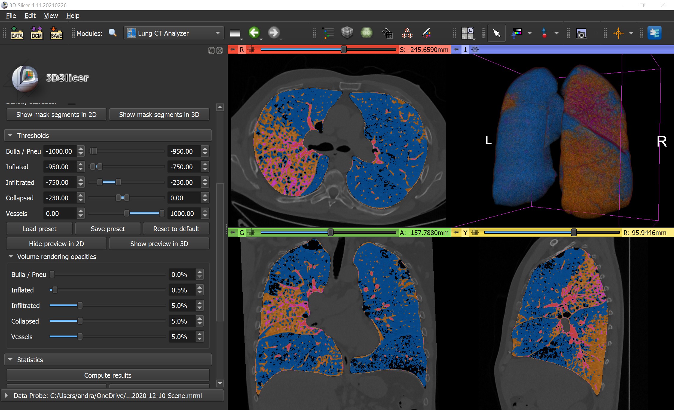
Lung CT analysis for COVID-19
LungCTAnalyzer extension offers automated lung segmentation and quantative analysis for COVID-19 cases. video > learn more >
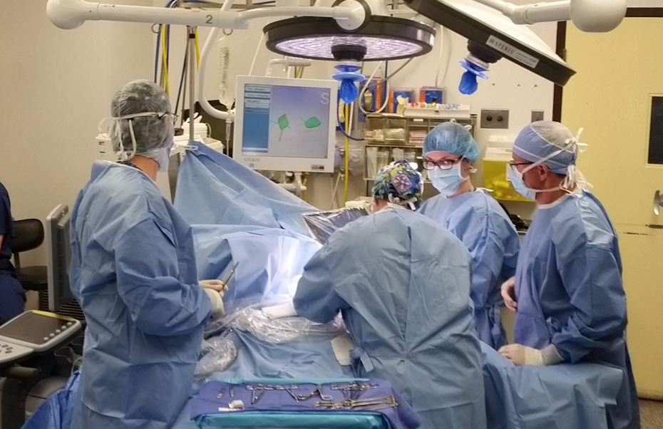
Surgical navigation
3D Slicer is used in real-time navigation of breast cancer surgery. video > journal article > learn more >
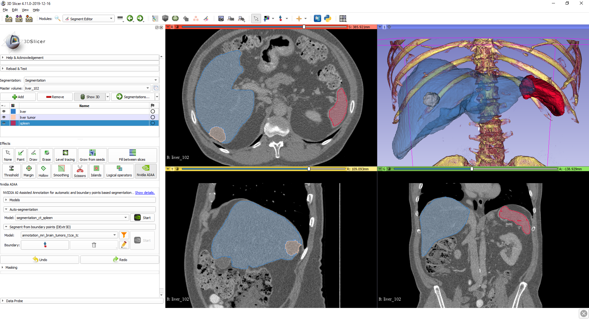
AI-assisted segmentation
AI-assisted annotation tools can automatically segment anatomical structures using pre-trained or custom models. See this tutorial video > or tutorial material > and
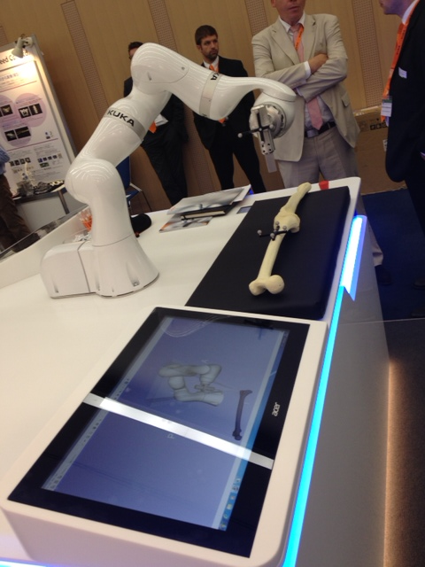
Robot-assisted interventions
Slicer is connected to a KUKA robot for visualization of 3D models of the robot, the anatomy, and the workspace. Demo at CARS 2014 in Fukuoka, Japan.The system is originally developed at NA-MIC Summer Project Week. learn more >
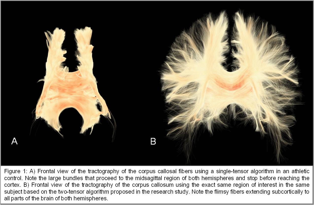
Tractography
UKFTractography is a module for computing tractography of DWI images using an unscented Kalman filter. Because of its 2-tensor algorithm, it is able to model fiber crossings and capture many more fibers than a single tensor algorithm. learn more >
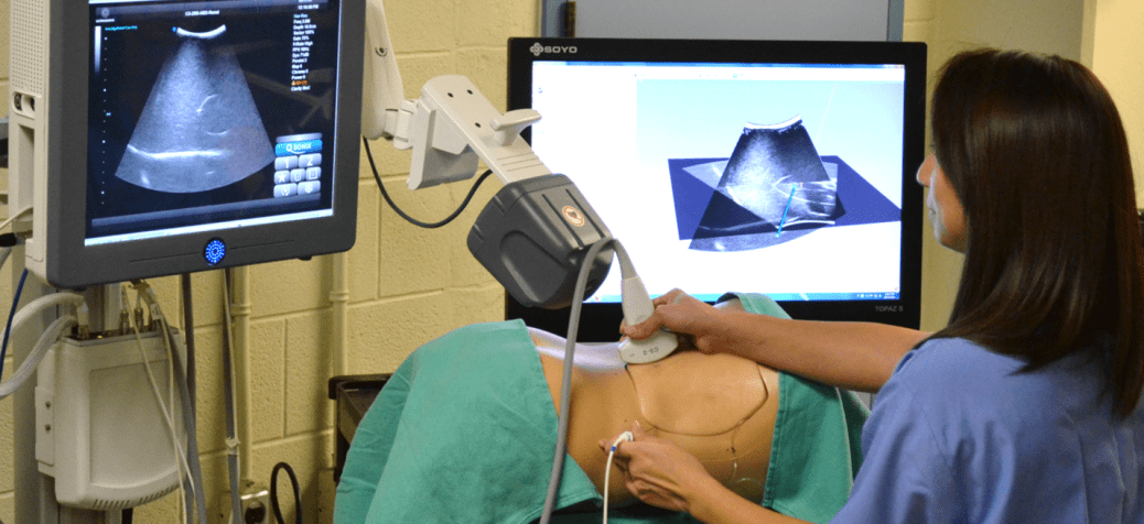
Tracked ultrasound for needle guidance
Tracked ultrasound snapshots enhance needle guidance for percutaneous renal access. video > learn more >
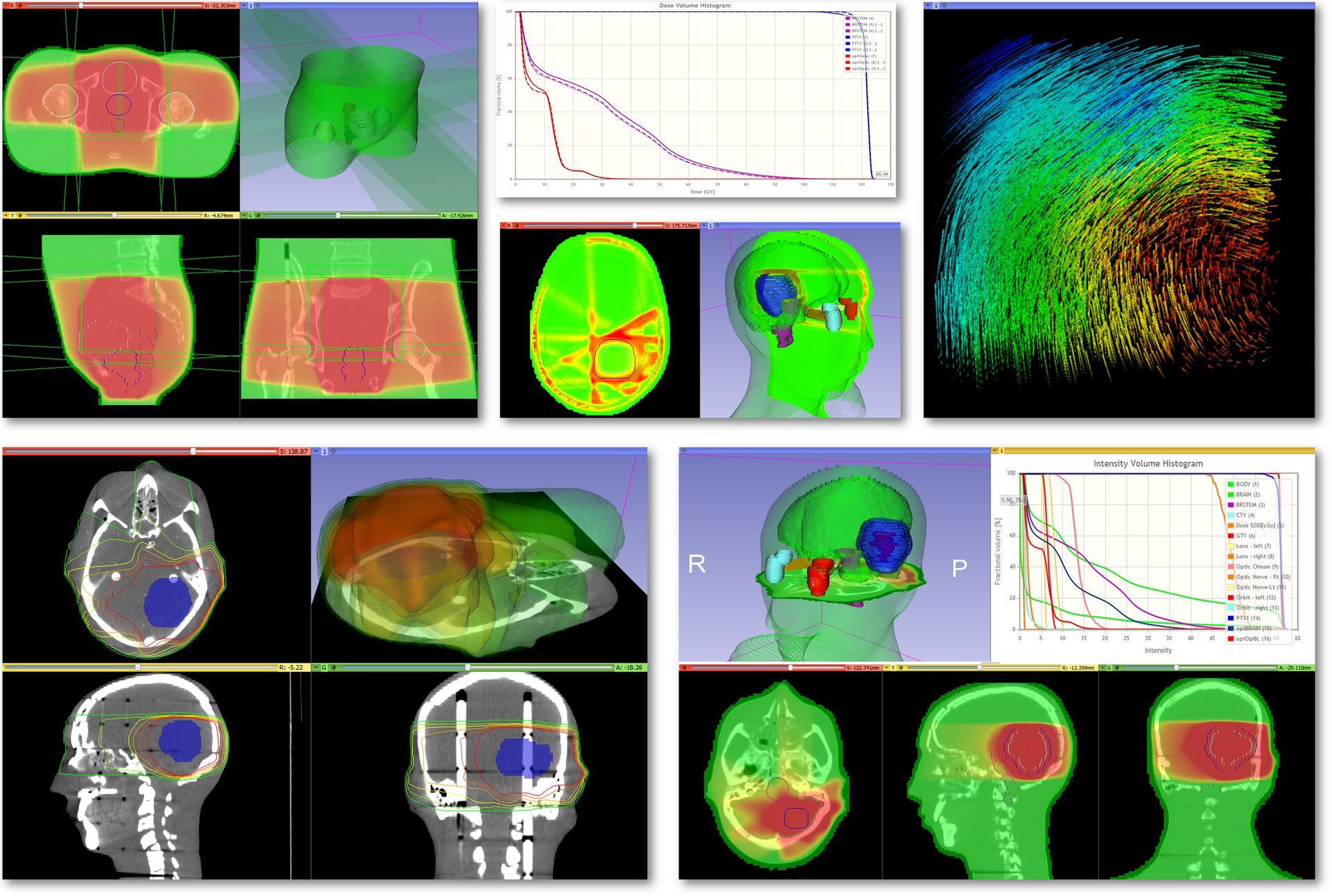
Adaptive radiation therapy
SlicerRT extension is a radiation therapy toolkit for 3D Slicer, containing DICOM RT import/export, visualization, and analysis. learn more >
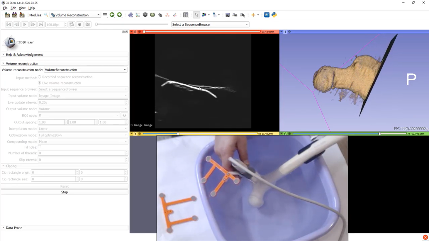
Real-time 3D ultrasound reconstruction
3D volume is reconstructed from real-time tracked ultrasound using SlicerIGT and SlicerIGSIO extensions video > learn more >
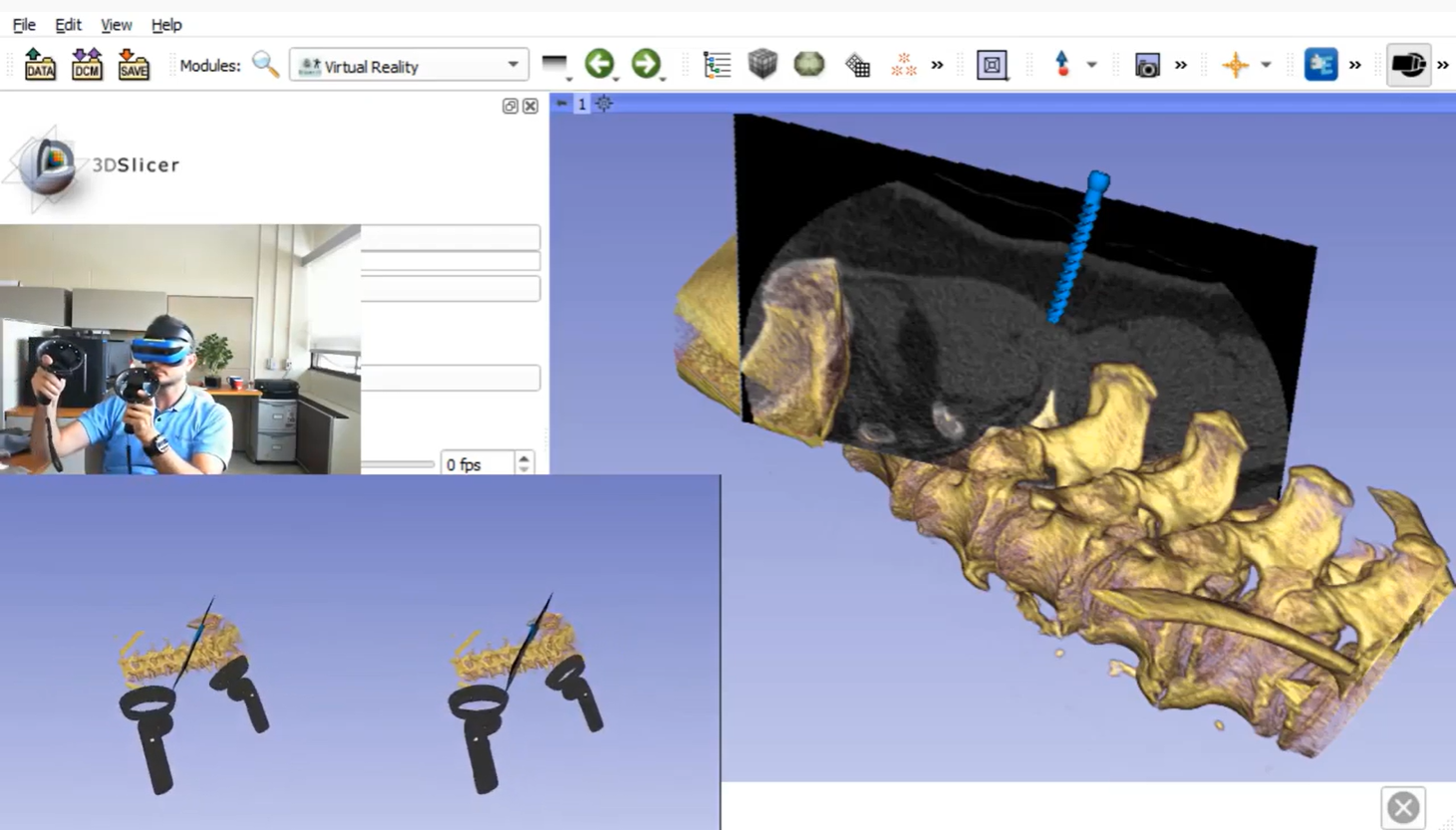
Virtual reality
Everything that is shown in the 3D view (volume rendering, real-time surgical navigation, tractography, etc.) can be displayed and interacted with in virtual reality using SlicerVR extension. video > learn more >
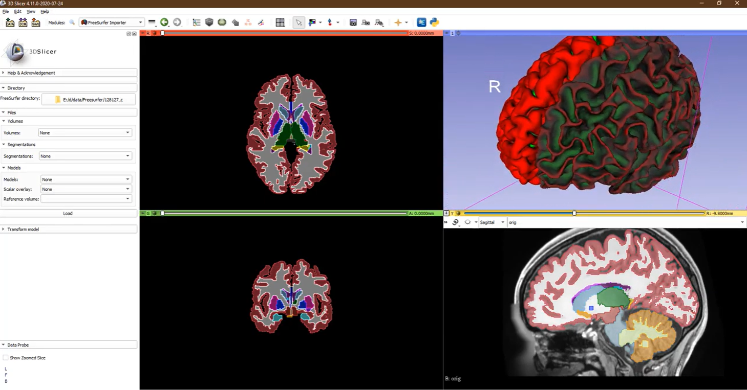
FreeSurfer support
Import volumes, segmentations, surfaces from FreeSurfer then edit and analyze them. video > learn more >
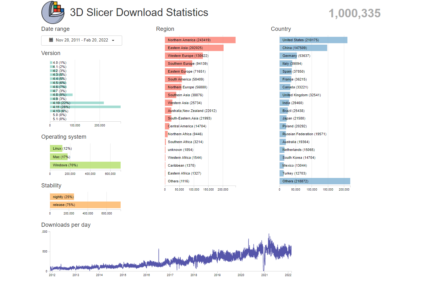
1 million downloads
3D Slicer application installers have been downloaded over 1 million times in the past 10 years. learn more >
 DICOM standard interoperability